
New aspects of high-resolution ultrasound for tumor detection and treatments: M-Elite Program
The concept of bringing together physicians with ultrasound expertise worldwide and ultrasound technology developers in terms of planning, design, and production would offer extensive opportunities for advancing ultrasound diagnostics. By taking part in initiatives based on this concept, physicians would have the chance to directly explore and evaluate the most up-to-date ultrasound technology for both diagnostic purposes and ultrasound-guided interventions during patient examinations at university ultrasound centres in China. The exchange of knowledge with fellow professionals and system developers would also be a significant component of this experience [1–11].
This concept was deliberated in small group discussions and individual meetings, focusing on forthcoming advancements in technology. Subsequently, there was an in-depth exploration of standards for scientific publications and environmentally conscious aspects of science and technology. Finally, an individual-focused survey was conducted to assess the potential for fostering comprehensive exchanges and the possibility of engaging in important, future-proof ultrasound developments via social media platforms, specifically through case discussions.
The prolonged duration of the pandemic has underscored the significance of direct interpersonal exchanges among ultrasound technology developers, investigators, users, learners, teachers, and researchers worldwide. Among this community, a notable segment actively participates in the M-Elite initiative, which facilitates on-site interactions at research and development centres across China. It is from these interactions that the current “M-Elite Program” format originated. Regarding the latest advancements in ultrasound technology, it is essential to conduct multi-centre evaluations on each imaging modality to assess their suitability for future clinical applications. This approach will allow for defining the threshold values for tissue properties (such as fibrosis and inflammation) in correlation with various diseases.
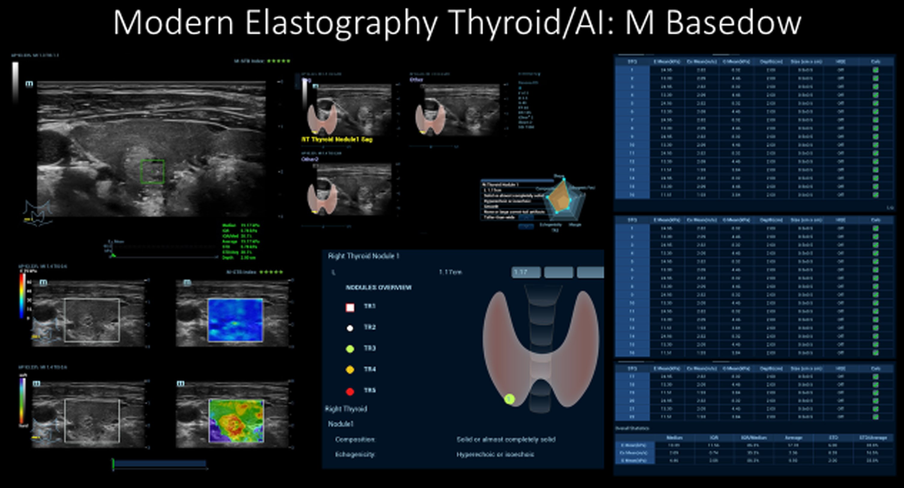
Recent elastography techniques, including Shear Wave, 2D, and STQ, liver texture analysis (LTI), and fat measurement through ultrasound attenuation analysis (USAT) enable comprehensive non-invasive parenchymal analyses using ultrasound. A variety of applications of contrast-enhanced ultrasonography (CEUS), combined with parametric imaging and perfusion analysis, which were previously performed in limited groups, provide a basis for better understanding changes in dynamic microvascularization down to the capillary level. Additionally, the integration of artificial intelligence (AI) can further refine the process and optimize reporting based on international standards (Fig. 1).
Fig. 1
Modern multimodal thyroid diagnostics with artificial intelligence (AI).
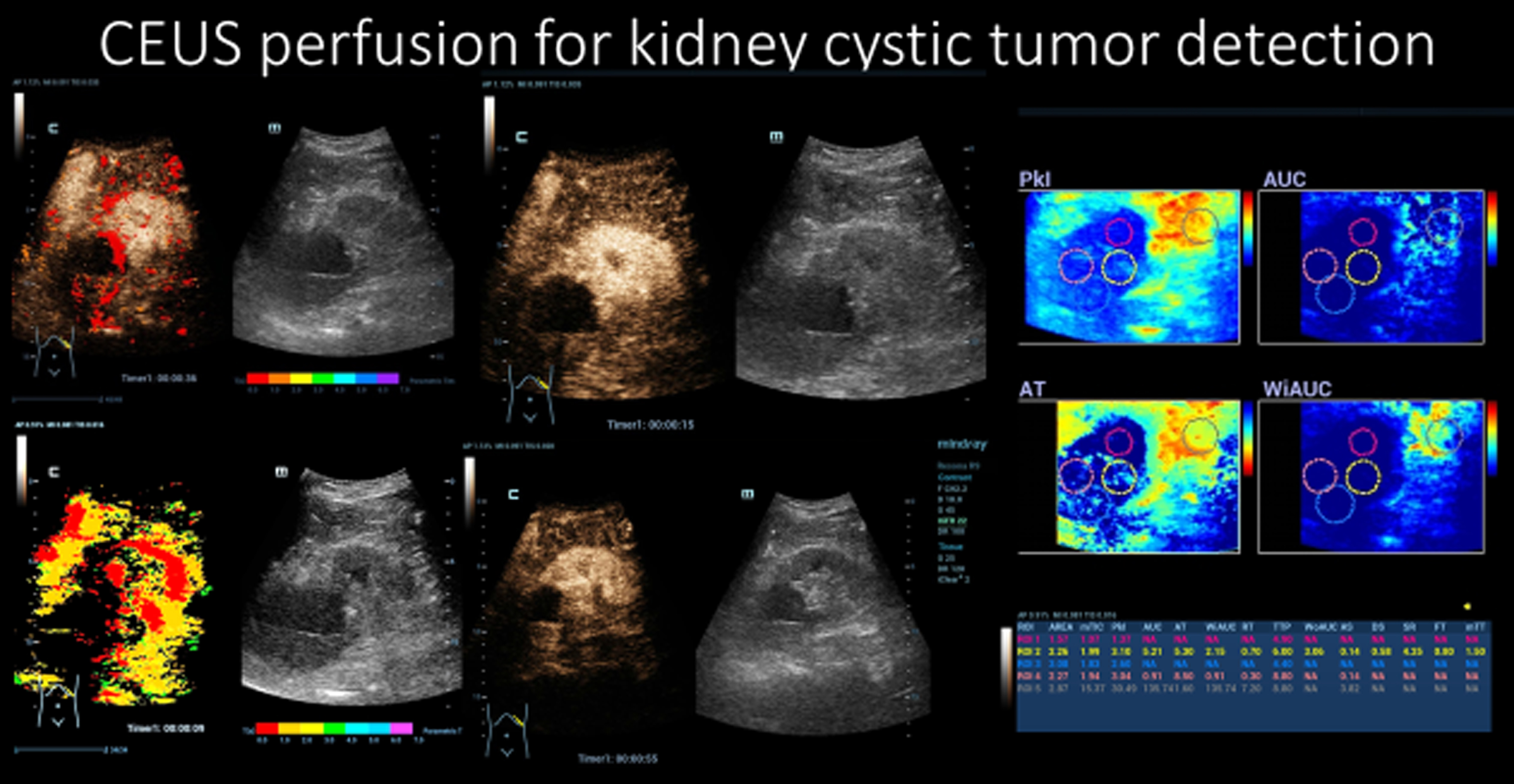
High-resolution ultrasound techniques, such as ZST and HiFR CEUS, offer significantly improved capabilities for early tumor detection and the ultrasound-guided treatment of tumors. In the future, fusion techniques will become increasingly accessible to enable the integration of different imaging modalities. Moreover, the utilization of 3D techniques with fusion and navigation will elevate surgical and interventional tumor treatments using ultrasound-guided robotics to unparalleled standards (Fig. 2).
Fig. 2
CEUS perfusion for the detection of a small intracystic kidney tumor.
Improved mobility, fast data transfer, exceptional resolution, and customizable examination interfaces and operator tools make ultrasound training and patient-related applications much easier, particularly in emergencies and challenging environments. This is the point at which M-Elite Expertise comes to the forefront and demonstrates its full potential.
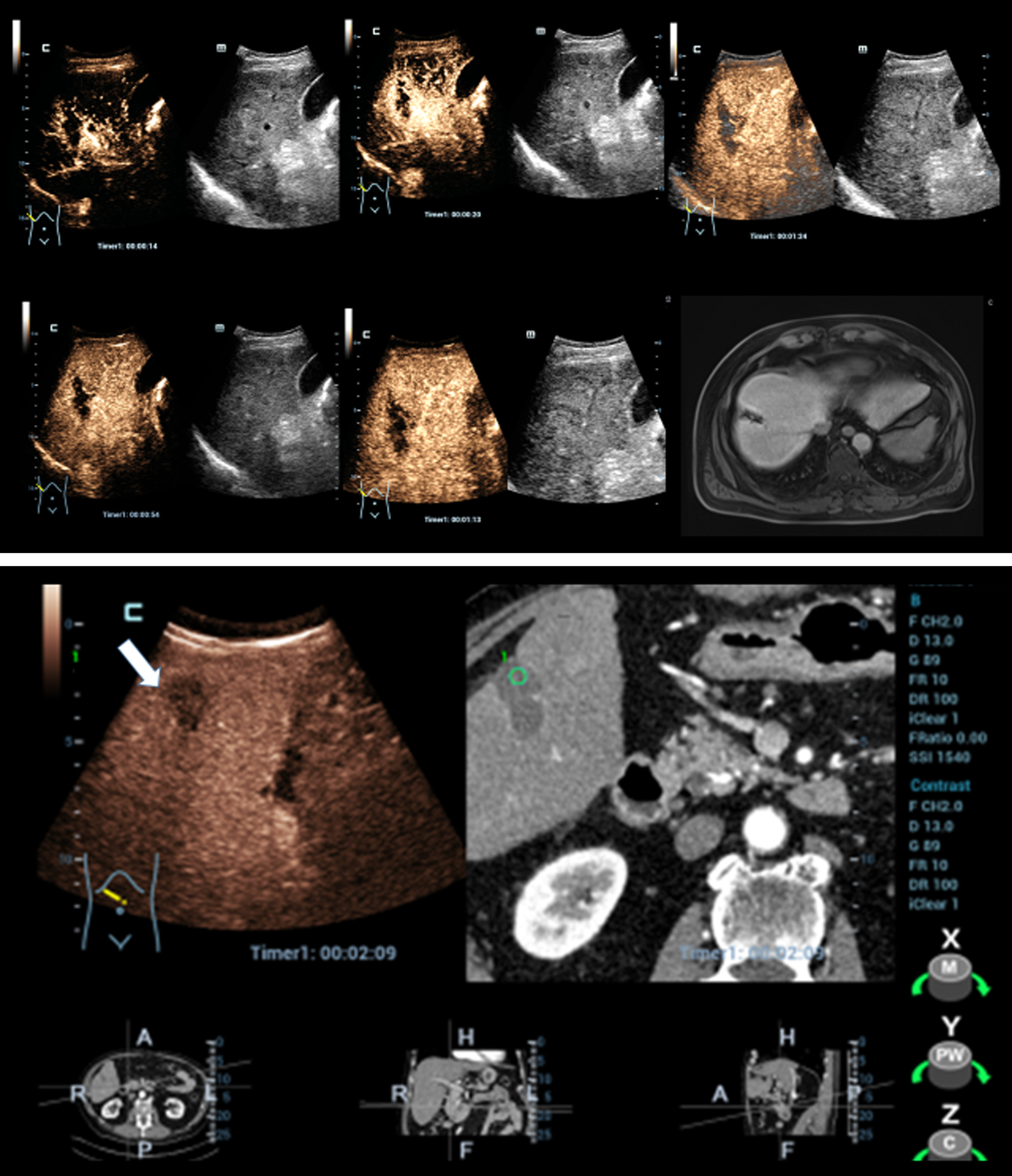
Cutting-edge ultrasound technology addresses the challenges posed by pandemics by offering fast and accurate imaging, maintaining superior hygiene standards, offering flexibility with extended battery life, and enabling more individualized treatments through advanced innovations (Fig. 3a/b).
Fig. 3
a/b: Dynamic CEUS using HiFR mode in correlation to contrast-enhanced MRI and fusion CEUS with contrast-enhanced CT to control successful microwave ablation (MWA) of a liver tumor with complete devascularization.
The combination of our latest research findings on CEUS parametric and perfusion analysis, with a focus on the diagnosis and interventions of liver tumors, thyroid tumors, lymph nodes, and breast tumors, alongside the implementation of modern elastography techniques and pioneering use of AI, has opened up avenues for conducting multicentre studies. Therefore, the M-Elite program serves as a foundation for developing effective educational concepts for students, sonographers, and university research projects.
The strong partnership between M-Elite and microcirculation departments establishes a solid groundwork for an innovative, technology-driven approach to ultrasound, state-of-the-art intensive monitoring, and enhanced laboratory diagnostics for comprehensive blood cell analysis.
This time, M-Elite carried out an exchange program at the First Affiliated Hospital of Sun Yat-Sen University (FAH-SUMS), whose medical ultrasound department is known as one of the top four ultrasound centres in China. They place great emphasis on the research of ultrasound medical treatments, educational training, and scientific investigation. Each year, the College of Medical Ultrasonic Medicine at FAH-SUMS offers a wide range of teaching and training programs for residents, graduate students, and studying physicians. Furthermore, they organize English-language teaching and exchange activities for the international community.
During the exchange program, we engaged in discussions covering various topics such as high-resolution elastic ultrasound, CEUS, interventional ultrasound, new parametric imaging, and ultrasound medical education. We also had the opportunity to gain insights into their ongoing projects and advancements in areas of single-mode fusion navigation, AI, and ultrasonic technology.
Conclusion
This exchange experience has strongly reinforced the notion that ultrasound transcends national boundaries, enabling seamless communication through visual images across the world. Ultrasound knows no bounds, as it offers potential solutions to numerous challenging issues encountered in clinical practice, fostering invention and innovation in its utilization. The possibilities within ultrasound technology are limitless, and the continuous development and implementation of new technologies have encouraged doctors from different countries to continue exploring advancements for the betterment of human health.
References
[1] | Daopeng Yang , Bowen Zhuang , Yan Wang, , et al. High-Frequency US for BK Polyomavirus-associated Nephropathy after Kidney Transplant. Radiology. (2022) ; 304: (2):333–41. |
[2] | Wenying Zhou , Yang Yang , Cheng Yu , et al. Ensembled deep learning model outperforms human experts in diagnosing biliary atresia from sonographic gallbladder images. Nat Commun. (2021) ;12: (1):1259. |
[3] | Fei Liu , Dan Liu , KunWang , et al. Deep Learning Radiomics Based on Contrast-Enhanced Ultrasound Might Optimize Curative Treatments for Very-Early or Early-Stage Hepatocellular Carcinoma Patients. Liver Cancer. (2020) ;9: (4):397–413. |
[4] | Zhang Xiaoer , Huang Guanglian , Ye Jieyi , et al. 3-D Contrast-Enhanced Ultrasound Fusion Imaging: A New Technique to Evaluate the Ablative Margin of Radiofrequency Ablation for Hepatocellular Carcinoma. Ultrasound Med Biol. (2019) ;45: (8):1933–43. |
[5] | Jung EM , Dong Y , Jung F . Current aspects of multimodal ultrasound liver diagnostics using contrast-enhanced ultrasonography (CEUS), fat evaluation, fibrosis assessment, and perfusion analysis - An update. Clin Hemorheol Microcirc. (2023) ;83: (2):181–93 doi: 10.3233/CH-239100. |
[6] | Jung EM , Stroszczynski C , Jung F Advanced multimodal imaging of solid thyroid lesions with artificial intelligence optimized B-mode, elastography, and contrast-enhanced ultrasonography parametric and with perfusion imaging: Initial results. Clin Hemorheol Microcirc. 2023. doi: 10.3233/CH-239102. Online ahead of print. |
[7] | Jung EM , Moran VO , Engel M , Krüger-Genge A , Stroszczynski C , Jung F . Modified contrast-enhanced ultrasonography with the new high-resolution examination technique of high frame rate contrast-enhanced ultrasound (HiFR-CEUS) for characterization of liver lesions: First results. Clin Hemorheol Microcirc. (2023) ;83: (1):31–46 doi:10.3233/CH-221449. |
[8] | Brandenstein M , Wiesinger I , Künzel J , Hornung M , Stroszczynski C , Jung EM . Multiparametric Sonographic Imaging of Thyroid Lesions: Chances of B-Mode, Elastography and CEUS in Relation to Preoperative Histopathology. Cancers (Basel). (2022) ;14: (19):4745. doi: 10.3390/cancers14194745. |
[9] | Qiu Y , Yang D , Zhang Q , Chen K , Dong Y , Wang WP . V Flow technology in measurement of wall shear stress of common carotid arteries in healthy adults: Feasibility and normal values. Clin Hemorheol Microcirc. (2020) ;74: (4):453–62. doi: 10.3233/CH-190719. |
[10] | Ding J , Du Y , Zhao R , Yang Q , Zhu L , Tong Y , Wen C , Wang M . Detection of Abnormal Wall Shear Stress and Oscillatory Shear Index via Ultrasound Vector Flow Imaging as Possible Indicators for Arteriovenous Fistula Stenosis in Hemodialysis. Ultrasound Med Biol. (2023) ;49: (8):1830–6. |
[11] | Zhu JE , Li JX , Zhang HL , Li XL , Sun LP , Yu SY , Xu HX . Sequential thermal ablation in combination with sclerotherapy using lauromacrogol as a successful translative therapy for an unresectable huge biliary cystadenocarcinoma: The first experience assisted by contrast-enhanced ultrasound. Clin Hemorheol Microcirc. (2022) ;82: (1):95–105. |


