Searching for just a few words should be enough to get started. If you need to make more complex queries, use the tips below to guide you.
Purchase individual online access for 1 year to this journal.
Price: EUR 210.00Impact Factor 2024: 1.7
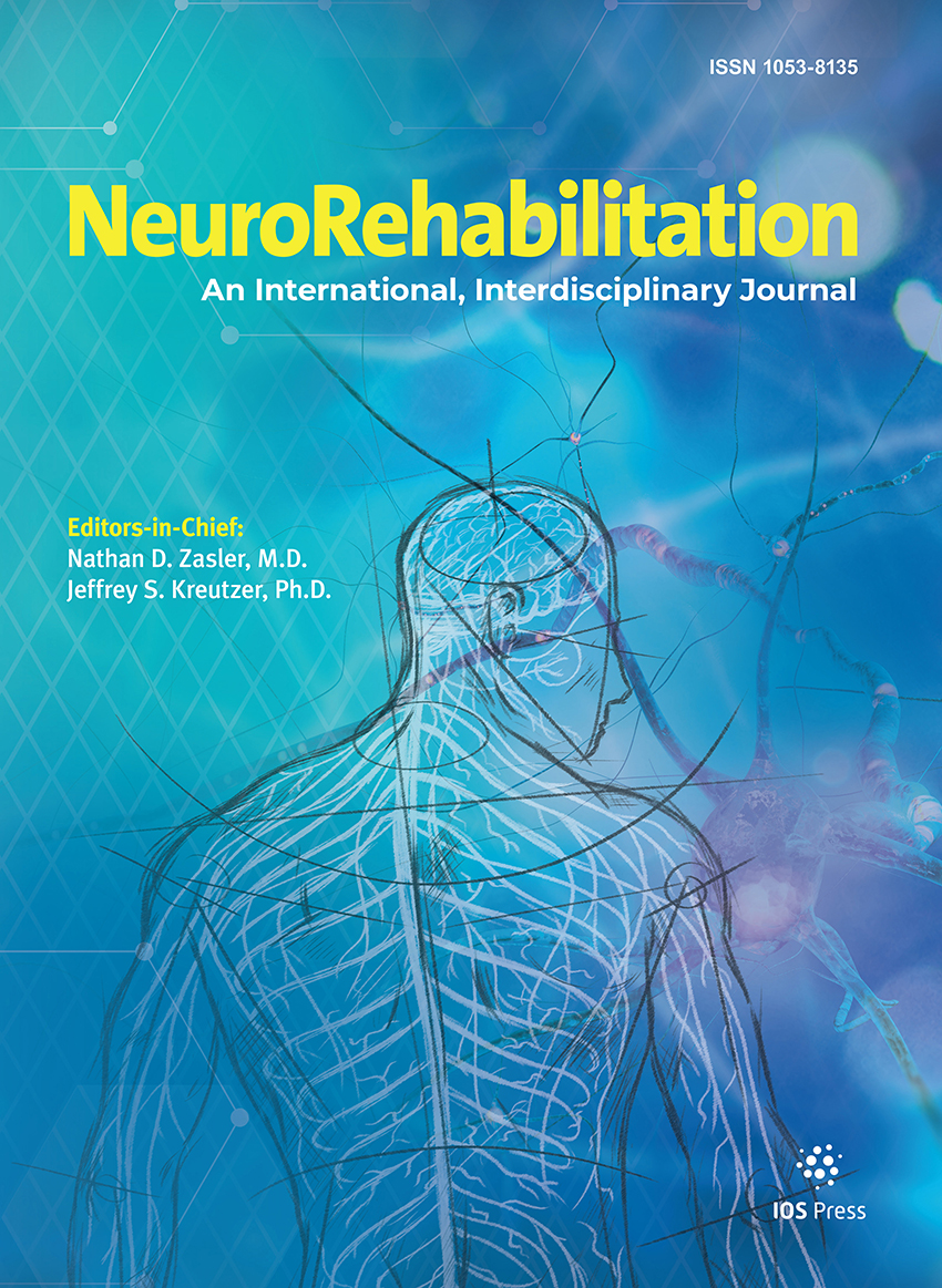
NeuroRehabilitation, an international, interdisciplinary, peer-reviewed journal, publishes manuscripts focused on scientifically based, practical information relevant to all aspects of neurologic rehabilitation. We publish unsolicited papers detailing original work/research that covers the full life span and range of neurological disabilities including stroke, spinal cord injury, traumatic brain injury, neuromuscular disease and other neurological disorders.
We also publish thematically organized issues that focus on specific clinical disorders, types of therapy and age groups. Proposals for thematic issues and suggestions for issue editors are welcomed.
Authors: Silverberg, Noah D. | Iverson, Grant L.
Article Type: Research Article
Abstract: In his seminal article, Physiogenesis and Psychogenesis in the ‘Post-Concussional Syndrome,’ Lishman (1988) proposed that neurobiological factors account for the development of the post-concussion syndrome and psychological factors become primarily responsible for maintaining it in the chronic phase. Over the 20 years that followed, researchers have advanced our understanding of the etiology of the post-concussion syndrome. Our review of this evidence suggests that neurobiological and psychological factors play a causal role in post-concussion symptoms from the outset, and thus, Lishman's causal model should be updated. If we can clinically identify individuals on a trajectory of poor recovery in the …acute post-injury stage, then we can direct secondary prevention towards modifiable risk factors. Show more
Keywords: Mild traumatic brain injury, etiology, rehabilitation
DOI: 10.3233/NRE-2011-0708
Citation: NeuroRehabilitation, vol. 29, no. 4, pp. 317-329, 2011
Authors: Tracy, Joseph I. | Osipowicz, Karol Z.
Article Type: Research Article
Abstract: Functional neuroimaging technologies are increasingly being used to predict cognitive/behavioral outcomes after the initiation of clinical interventions such as resective surgery or cognitive rehabilitation. We provide a conceptual model and a case example to explain how the results from various neuroimaging techniques can be integrated to answer important questions about clinical recovery such as whether neural reorganization has occurred and, if so, the type of adaptive cognitive mechanism driving this reorganization. This proposed framework and its use in interpreting neuroimaging outcomes studies should help uncover the principles that govern neural reorganization, and be of use to any patient for whom …the risk, or potential benefit, of brain-based interventions is unknown. Show more
Keywords: Neural reorganization, neuroplasticity, cognitive mechanism, cognitive reorganization, brain recovery
DOI: 10.3233/NRE-2011-0709
Citation: NeuroRehabilitation, vol. 29, no. 4, pp. 331-338, 2011
Authors: Jang, Sung Ho
Article Type: Research Article
Abstract: The corticospinal tract (CST) is the most important neural tract for motor function in the human brain. Therefore, clarification of CST injury would be an important topic in traumatic brain injury (TBI) rehabilitation. In this review, I reviewed diffusion tensor imaging (DTI) studies on CST injuries in terms of etiology and recovery in patients with TBI. Although DTI has several unique advantages for research on CST injury in TBI, only a dozen DTI studies on this topic have been reported: etiology of CST injury (9 studies), recovery of CST injury (3 studies). As for the etiology of CST injury in …TBI, the previous studies have demonstrated the usefulness of DTI in diagnosis of CST injury in cases of diffuse axonal injury, transtentorial herniation, cerebral hemorrhage, and cortical contusion; moreover, according to the severity of TBI. The three studies on recovery of CST injury focused on recovery of a CST injured by diffuse axonal injury. In the future, we suggest an increase in the total number of DTI studies on this topic. In particular, research on recovery of CST injury should be encouraged. Moreover, studies of the various recovery mechanisms related to the CST are necessary. Show more
Keywords: Traumatic brain injury, diffuse axonal injury, traumatic axonal injury, diffusion tensor imaging, diffusion tensor tractography, motor recovery, corticospinal tract
DOI: 10.3233/NRE-2011-0710
Citation: NeuroRehabilitation, vol. 29, no. 4, pp. 339-345, 2011
Authors: Bakheit, A.M.O. | Fletcher, K. | Brennan, A.
Article Type: Research Article
Abstract: Abulia is a disorder of the executive and frontal lobe function. It is characterised by severe psychomotor slowing that is not due to depressive illness or catatonic schizophrenia. Abulia is thought to be due to disruption of the meso-cortico-limbic dopaminergic system. Preliminary evidence suggests that patients with abulia may respond to treatment with dopaminergic drugs. We extend this evidence by reporting a significant and sustained functional improvement in a severely abulic patient after treatment with co-beneldopa (Madopar).
Keywords: Abulia treatment, akinetic mutism, levodopa therapy
DOI: 10.3233/NRE-2011-0711
Citation: NeuroRehabilitation, vol. 29, no. 4, pp. 347-351, 2011
Authors: Giner Pascual, Manuel | Sebastià Alcácer, Vicente | Perez Pomares, Maria Victoria | Alcanyis-Alberola, Modesto
Article Type: Case Report
Abstract: Study Design: Case report. Objective: To describe a patient presenting with Brown-Séquard-plus syndrome treated in a conservative manner and to discuss the possible physiopathological mechanisms causing the injury. Methods: The case study of a 35-year-old woman who entered the hospital with a knife that had penetrated her neck through the left upper thoracic aperture and with a rising, back, right oblique trajectory. This patient developed Brown-Séquard-plus syndrome on the right side of her body. Results: The initial computerized tomography (CT) demonstrated that the tip of the knife was inside the right C7 vertebral foramen, …which not dissected the vertebral artery. The initial magnetic resonance imaging (MRI) and the MRI done 3 weeks later showed the presence of spinal cord ischemia on the right side at the C6-C7 level. This spinal cord ischemia was most likely caused after a vessel spasm of the vertebral artery. After conservative treatment, the patient evolved from a C rating on the ASIA scale to a D rating. Conclusion: In our department, spinal cord injuries after stab wounds are very rare, and they usually cause incomplete lesions that eventually lead to Brown-Séquard syndrome. In our patient, the spinal cord injury was due to a vasospasm of the vertebral artery, which was accompanied by good functional prognosis. MRI helped to define the physiopathologic mechanism of the injury and guided the appropriate treatment decision. Show more
Keywords: Brown-Séquard syndrome, stab injury, MRI
DOI: 10.3233/NRE-2011-0712
Citation: NeuroRehabilitation, vol. 29, no. 4, pp. 353-357, 2011
Authors: Lee, Mi Young | Jang, Sung Ho
Article Type: Research Article
Abstract: Many studies have reported that stroke patients can be accompanied by motor deficit of the unaffected extremities as well as the affected extremities. This suggests that neural control of motor function of unaffected extremities might be changed following stroke. However, very little is known about this topic. Using functional MRI (fMRI), we investigated changes in neural control of motor function of the unaffected hand in hemiparetic patients with cerebral infarct. Thirty-five hemiparetic stroke patients were recruited for this study. fMRI was performed at 1.5T during either affected or unaffected hand flexion-extension movements. We evaluated motor function of the affected upper …extremity using the upper Motricity index (UMI) and the medical research council (MRC) scale for finger extensor. From fMRI, LI (laterality index) was calculated for assessment of relative activity in the ipsilateral versus the contralateral primary sensorimotor cortex. Positive correlation between LIs was observed during affected and unaffected hand movements (r = 0.670, p = 0.000). LI of unaffected hand movements was also correlated with the affected UMI (r = 0.408, p = 0.015) and MRC of the affected hand extensor (r = 0.362, p = 0.033). We demonstrated that the ipsilateral (affected) motor cortex was recruited by unaffected hand movements in proportion to poor motor function of the affected upper extremity. Show more
Keywords: Functional MRI, hemiparesis, ipsilateral motor cortex, stroke
DOI: 10.3233/NRE-2011-0713
Citation: NeuroRehabilitation, vol. 29, no. 4, pp. 359-364, 2011
Authors: Kakuda, Wataru | Abo, Masahiro | Kobayashi, Kazushige | Momosaki, Ryo | Yokoi, Aki | Fukuda, Akiko | Umemori, Takuma
Article Type: Research Article
Abstract: Introduction: The purpose of this study was to clarify the safety, feasibility and efficacy of 6-Hz primed low-frequency repetitive transcranial magnetic stimulation (rTMS) applied with intensive occupational therapy (OT) for upper limb hemiparesis after stroke. Subjects and methods: Eleven patients with history of stroke and upper limb hemiparesis (age at intervention: 61.0 ± 13.7 years, time after stroke onset: 70.2 ± 39.8 months) were studied. Each patient received 22 sessions of 6-Hz primed low-frequency rTMS (10-min 6-Hz priming stimulation followed by 20-min low-frequency rTMS of 1-Hz) applied to the non-lesional hemisphere plus intensive OT comprising 60-min one-to-one training …and 60-min self-training during 15-day hospitalization. The motor function of the affected upper limb was evaluated by Fugl-Meyer Assessment (FMA) and Wolf Motor Function Test (WMFT) on the days of admission and discharge. Results: All patients completed the 15-day protocol without any adverse effects. The treatment increased the FMA score (from 42.2 ± 6.9 to 45.6 ± 7.2 points, p < 0.005) and shortened the log performance time of WMFT (from 3.26 ± 1.21 to 2.81 ± 1.26 sec, p < 0.05). Conclusions: The 15-day protocol of 6-Hz primed low-frequency rTMS combined with intensive OT seems safe and a potentially useful therapeutic modality for upper limb hemiparesis after stroke. Show more
Keywords: Stroke, upper limb hemiparesis, transcranial magnetic stimulation, occupational therapy
DOI: 10.3233/NRE-2011-0714
Citation: NeuroRehabilitation, vol. 29, no. 4, pp. 365-371, 2011
Authors: MahmoudAliloo, M. | Bakhshipour, A. | Hashemi, T. | Roofigari, A.R. | Hassan-Zadeh, Roghieh
Article Type: Research Article
Abstract: Introduction: The aim of this study is to determine the correlation of cognitive ability with functional sensibility. Method: 130 patients with median and ulnar nerve repair at the distal forearm level and wrist level were included. Mean time since surgery and age were 44.35 months (range 23–68) and, 33.44 years (range 18–71), respectively. The patients underwent an assessment of sensory function of the hand and a battery of specific tests for cognitive capacity. Results: A multiple linear regression analysis demonstrated a significant correlation between functional sensibility and cognitive capacity [Block design test (Beta = 0.40, p …= 0.00); Reaction time (Beta = −0.32, p = 0.00); The Stroop color word test (Beta = 0.16, p = 0.04)]. Discussion: The results suggested that, cognitive capacity factors are associated with functional sensibility after nerve repair. These results may be mentioned cognitive rehabilitation programs would enhanced functional outcome following nerve repair. Show more
Keywords: Cognitive capacity, functional sensibility, nerve repair, upper extremity
DOI: 10.3233/NRE-2011-0715
Citation: NeuroRehabilitation, vol. 29, no. 4, pp. 373-379, 2011
Authors: Rodríguez-Mutuberría, Liván | Álvarez-González, Lázaro | López, Maikel | Bender-del Busto, Juan E. | Fernández-Martínez, Elizabeth | Martínez-Segón, Susana | Bergado, Jorge A.
Article Type: Research Article
Abstract: The present paper shows the result of an open prospective study performed to evaluate the tolerance and efficacy of a program for neurological restoration (PRN) in stroke patients. The PRN is organized 4 weeks cycles – 39 hours per week – and applied by a team of physical, occupational, and speech therapists, physiatrists, psychologists, clinicians and nurses; directed by a neurologist. The first phase of treatment aims to increase the physical capacity and tolerance to exercise. The second phase trains specific abilities (balance, posture, gait and handling). Drugs were only used to modulate physical or mood disorders, spasticity, or pain. …The study was performed in 80 stroke patients attended in our institution (2005–2007). Only patients with a confirmed diagnosis of stroke in the carotid territories, over 15 years old, and not least than 6 months post-ictal evolution were included. Tolerance to treatment was very good, with only 4 adverse events not related to treatment. The neurological condition was evaluated using the Scandinavian Stroke Scale (SSS), and the functional condition using the Barthel Index (BI). The results show significant improvements both in the neurological (113.45 ± 1.59%) and functional (130.11 ± 5.17%) condition after one treatment cycle, which improved further when therapy continued for a second cycle (233.71 ± 7.76% and 207.62 ± 27.16% respectively). Severity of the impairment was not a negative predictor of the outcome. Age correlated negatively with the initial condition, but does not prevent improvement. Sex, time of evolution, affected hemisphere or interactions among them did not influence the outcome. These results demonstrate that the PRN is well tolerated and effective promoting recovery even in chronic stroke patients. Show more
Keywords: Neurological restoration, neurorehabilitation, stroke
DOI: 10.3233/NRE-2011-0716
Citation: NeuroRehabilitation, vol. 29, no. 4, pp. 381-391, 2011
Authors: Sabut, Sukanta K. | Sikdar, Chhanda | Kumar, Ratnesh | Mahadevappa, Manjunatha
Article Type: Research Article
Abstract: Objective: To evaluate the therapeutic effects of Functional Electrical Stimulation (FES) of the tibialis anterior muscle on plantarflexor spasticity, dorsiflexor strength, voluntary ankle dorsiflexion, and lower extremity motor recovery with stroke survivors. Design: We conducted a prospective interventional study. Setting: Rehabilitation ward, physiotherapy unit and gait analysis laboratory. Participants: Fifty-one patients with foot drop resulting from stroke. Intervention: The functional electrical stimulation (FES) group (n = 27) received 20–30 minutes of electrical stimulation to the peroneal nerve and anterior tibial muscle of the paretic limb along with conventional rehabilitation program (CRP). …The control group (n = 24) treated with CRP only. The subjects were treated 1 hr per day, 5 days a week, for 12 weeks. Main outcome measures: Plantarflexor spasticity measured by modified ashworth scale (MAS), dorsiflexion strength measured by manual muscle test (MMT), active/passive ankle joint dorsiflexion range of motion, and lower-extremity motor recovery by Fugl-Meyer assessment (FMA) scale. Results: After 12 weeks of treatment, there was a significant reduction in a plantarflexor spasticity by 38.3% in the FES group and 21.2% in control group (P < 0.05), between the beginning and end of the trial. Dorsiflexor muscle strength was increased significantly by 56.6% and 27.7% in the FES group and control group, respectively. Similarly, voluntary ankle dorsiflexion and lower-extremity motor function improved significantly in both the groups. No significant differences were found in the baseline measurements among groups. When compared with control group, a significant improvement (p < 0.05) was measured in all assessed parameters in the FES group at post-treatment assessment, thus FES therapy has better effect on recovery process in post-stroke rehabilitation. Conclusions: Therapy combining FES and conventional rehabilitation program was superior to a conventional rehabilitation program alone, in terms of reducing spasticity, improving dorsiflexor strength and lower extremity motor recovery in stroke patients. Show more
Keywords: Stroke, electrical stimulation, spasticity, foot-drop, motor recovery
DOI: 10.3233/NRE-2011-0717
Citation: NeuroRehabilitation, vol. 29, no. 4, pp. 393-400, 2011
Authors: Kwon, Hyeok Gyu | Jang, Sung Ho
Article Type: Research Article
Abstract: The arcuate fasciculus (AF) is the neural tract that connects Wernicke's area and Broca's area. The main role of the AF is speech repetition; therefore, injury to the AF typically causes conduction aphasia. We report on a patient who showed excellent recovery of aphasia despite complete injury of the AF due to a cerebral infarct. A 54-year-old, right-handed male presented with aphasia and right hemiparesis. Brain MRI showed an infarct in the left centrum semiovale and corona radiata. Diffusion tensor tractography for the AF was reconstructed using DTI-studio software. The Korean-Western Aphasia Battery (K-WAB) was used for measurement of language …function. On K-WAB at 1 week after onset, his aphasia type was compatible with global aphasia (aphasia quotient: 12‰, fluency: 5‰, comprehension: 24‰, repetition: 15‰, and naming: 31‰). The patient underwent rehabilitative therapy, including language therapy and medication, which is known to facilitate recovery from aphasia, for a period of 24 months. His aphasia had improved to a nearly normal state at 30 months after onset; aphasia quotient: 93‰ (fluency: 91‰, comprehension: 92‰, repetition: 85‰, and naming: 96‰). The left AF showed a complete disruption on 27-month diffusion tensor tractography. Findings from this study suggest the possibility that aphasia might show good recovery, even in cases of severe injury of the AF. Show more
Keywords: Diffusion tensor imaging, diffusion tensor tractography, stroke, arcuate fasciculus, aphasia
DOI: 10.3233/NRE-2011-0718
Citation: NeuroRehabilitation, vol. 29, no. 4, pp. 401-404, 2011
Authors: Folland, Jonathan P. | Haas, Bernhard | Castle, Paul C.
Article Type: Research Article
Abstract: The specific neuromuscular mechanisms for compromised muscle strength with PD, and the improvement that occurs with medication, have not been clearly delineated. This study assessed knee extension and flexion strength of PD patients whilst on and off medication and examined the neural mechanisms responsible for any changes. Ten idiopathic PD patients were assessed whilst on and off medication (⩾ 12-h after drug withdrawal), ∼ 7 days apart. Isometric strength of the knee extensors and flexors was assessed, and the interpolated twitch technique used to measure activation of the knee extensors. Surface EMG was also used to measure neural drive to …the agonists and antagonists. Without medication isometric strength of the knee extensors (7%) and flexors (11%) was impaired and the interpolated twitch technique revealed activation of the knee extensors was reduced (8%, P = 0.005). Maximum agonist amplitudes for nkee extension and flexion were unchanged off-medication (0.59 < P < 0.77). The agonist and antagonist EMG-force relationships, and the maximum antagonist EMG, were unaffected by medication withdrawal. The decrease in knee extension strength when PD patients were off medication was due to reduced activation of the agonist muscle, rather than any change in antagonist co-activation, and these changes were associated with reduced locomotory performance. Show more
Keywords: Parkinson's disease, muscle strength, medication, activation, co-activation, interpolated twitch technique
DOI: 10.3233/NRE-2011-0719
Citation: NeuroRehabilitation, vol. 29, no. 4, pp. 405-411, 2011
Authors: Vidal, Xavier | Morral, Antonio | Costa, Lluís | Tur, Miriam
Article Type: Research Article
Abstract: Aim: The aim of this study was to evaluate the efficacy and safety of radial extracorporeal shock wave therapy (rESWT) in the treatment of spasticity in patients with cerebral palsy. Methods: Fifteen patients with spastic cerebral palsy, 12 men and 3 women, aged 10–46 years (mean age 31). The 15 patients presented 40 spastic muscles that were divided in three groups using a computerized random-number generator. The first group, received rESWT in spastic muscle. The second group received rESWT in spastic muscle + rESWT in antagonist muscle. The third group received placebo. Range of motion and Ashworth Scale …were performed. This study is a randomized, placebo-controlled clinical trial. The patients were treated in 3 sessions at intervals of one week. Results: There are significant differences between groups treated with rESWT and group placebo. A significant decrease in the Ashworth Scale, an increase in the range of motion, were observed in all patients that were treated with rESWT. Positive results were maintained for at least 2 months after treatment. Interpretation: The treatment with rESWT is more effective than placebo in decreasing spasticity of patients with CP. Show more
Keywords: Cerebral palsy, spasticity, extracorporeal shock wave therapy, hypertonia, muscle tone
DOI: 10.3233/NRE-2011-0720
Citation: NeuroRehabilitation, vol. 29, no. 4, pp. 413-419, 2011
Article Type: Other
DOI: 10.3233/NRE-2011-29414
Citation: NeuroRehabilitation, vol. 29, no. 4, pp. 421-424, 2011
Article Type: Other
DOI: 10.3233/NRE-2011-29415
Citation: NeuroRehabilitation, vol. 29, no. 4, pp. 425-426, 2011
IOS Press, Inc.
6751 Tepper Drive
Clifton, VA 20124
USA
Tel: +1 703 830 6300
Fax: +1 703 830 2300
[email protected]
For editorial issues, like the status of your submitted paper or proposals, write to [email protected]
IOS Press
Nieuwe Hemweg 6B
1013 BG Amsterdam
The Netherlands
Tel: +31 20 688 3355
Fax: +31 20 687 0091
[email protected]
For editorial issues, permissions, book requests, submissions and proceedings, contact the Amsterdam office [email protected]
Inspirees International (China Office)
Ciyunsi Beili 207(CapitaLand), Bld 1, 7-901
100025, Beijing
China
Free service line: 400 661 8717
Fax: +86 10 8446 7947
[email protected]
For editorial issues, like the status of your submitted paper or proposals, write to [email protected]
如果您在出版方面需要帮助或有任何建, 件至: [email protected]