
Role of non-invasive objective markers for the rehabilitative diagnosis of central sensitization in patients with fibromyalgia: A systematic review
Abstract
BACKGROUND:
Central sensitization cannot be demonstrated directly in humans. Therefore, studies used different proxy markers (signs, symptoms and tools) to identify factors assumed to relate to central sensitization in humans, that is, Human Assumed Central Sensitization (HACS). The aims of this systematic review were to identify non-invasive objective markers of HACS and the instruments to assess these markers in patients with fibromyalgia (FM).
METHODS:
A systematic review was conducted with the following inclusion criteria: (1) adults, (2) diagnosed with FM, and (3) markers and instruments for HACS had to be non-invasive. Data were subsequently extracted, and studies were assessed for risk of bias using the quality assessment tools developed by the National Institute of Health.
RESULTS:
78 studies (
CONCLUSIONS:
This review provides an overview of non-invasive markers and instruments for the assessment of HACS in patients with FM. Implementing these findings into clinical settings may help to identify HACS in patients with FM.
1.Introduction
The term nociplastic pain is defined as “pain that arises from altered nociception despite no clear evidence of actual or threatened tissue damage causing the activation of peripheral nociceptors or evidence for disease or lesion of the somatosensory system causing the pai” [1]. Central sensitization (CS) can be described as “an increased responsiveness of nociceptive neurons in the central nervous system to their normal or subthreshold afferent input” and can therefore be an expression of nociplastic pain [2]. CS results in an enhanced nociceptive neural signaling, meaning that the stimulus intensity necessary to elicit the pain response is lowered, resulting in pain hypersensitivity [3]. This alteration in sensory processing systems is observed in animal experiments [4], and this phenomenon is supposed to be of value to explain multiple chronic pain conditions such as low back pain, osteoarthritis, temporomandibular disorders and fibromyalgia (FM) [5]. FM, with a worldwide prevalence of 2–4%, and temporomandibular disorders are among the most common causes of pain and disability related to CS [6]. Both disorder share features and are influenced by genetic, biological, and psychosocial factors, such as diet, obesity and stressful events [6, 7, 8]. Because there is no gold standard for the assessment of CS, the presence of it cannot be demonstrated directly in humans. Instead, studies used different proxy markers (signs, symptoms and tools) to identify factors assumed to relate to CS in human, that is, Human Assumed Central Sensitization (HACS). A proxy marker for HACS can be defined as an indirect measurable indicator of the assumed presence of CS. These proxy markers will further be referred to as ‘marker’ in this review. The term HACS has previously been defined in a review on HACS in patients with chronic low back pain [9]. As FM is related to CS, rehabilitative therapy could play a useful role in the improvement of pain-related and mobility symptoms [7]. Thus, improving the accuracy of FM diagnosis can aid in the rehabilitation process of patients with FM.
Because of the absence of a typical physiological abnormality specific to FM, the diagnosis of FM is based on clinical presentation only, with a diagnostic criterium using a list of eighteen body sites and experiencing pain in at least 11 of the 18 tender points [10]. The 1990 American College of Rheumatology (ACR) classification for diagnosis comprises the assessment of pain in eighteen body sites combined with the average scores of a self-administered questionnaire [11]. The revised 2010 ACR classification includes a calculation of the widespread pain index, a symptom severity scale and does not contain a tender point examination. Diagnostic studies in patients with FM were conducted with the aim of identifying HACS markers, varying from cerebrospinal fluid (CSF) and serum concentrations [12, 13, 14] to urinary metabolites such as creatine [11]. Simple clinical tests to objectively identify HACS markers may, however, contribute to setting more suitable and objective diagnosis which are clinically feasible. While there appear to be several studies available, an overview of the current state is missing. The aim of this study was to review the literature to determine non-invasive markers for the presence of HACS in patients with FM and the instruments needed for the assessment of these markers.
Table 1
Eligibility criteria for study selection
| Inclusion criteria | Exclusion criteria | |
|---|---|---|
| Population |
|
|
| Target condition |
|
|
| Type of studies |
|
|
| Outcomes |
|
|
2.Methods
The search strategy started with a broad search regarding non-invasive markers of HACS for three chronic musculoskeletal pain diagnoses: fibromyalgia, chronic low back pain and osteoarthritis. Due to the vast amount of hits provided by the search, the authors decided to split it in three parts. Therefore, the current study constitutes the first part of a larger review about pain processing in chronic musculoskeletal pain disorders and is focused on markers for HACS in patients with FM. A second part is focused on HACS in patients with chronic low back pain and a third study focusses on HACS in patients with osteoarthritis and other painful syndromes. The current systematic review was reported based on the Preferred Reporting Items for Systematic Reviews and Meta-Analyses (PRISMA) [15] and has been prospectively registered in Prospero (October 2020: CRD42020172382).
2.1Search strategy
Three electronic databases (PubMed, EMBASE and PsycINFO) were searched on 01/04/2022. MeSH terms in PubMed were incorporated in the search string. Keywords were divided into the three following categories: the target population consisted of patients with “Musculoskeletal Pain” OR “Chronic Pain”. The target condition was HACS. Because there is no consensus or uniformity in terminology, we used the following search terms for HACS conditions: “Central Sensitizatio” OR “Centralized Pain”, “Hypersensitivity” and synonyms. Finally, the outcome measures “Neurophysiological Biomarker” were related to non-invasive HACS markers. Non-invasive markers are defined as markers determined through a procedure that does not cause a break in the skin, nor creates contact with the mucosa or an internal body cavity. Synonyms of the different keyword groups constituted the search request. The entire search string is presented in Appendix A.
2.2Eligibility criteria
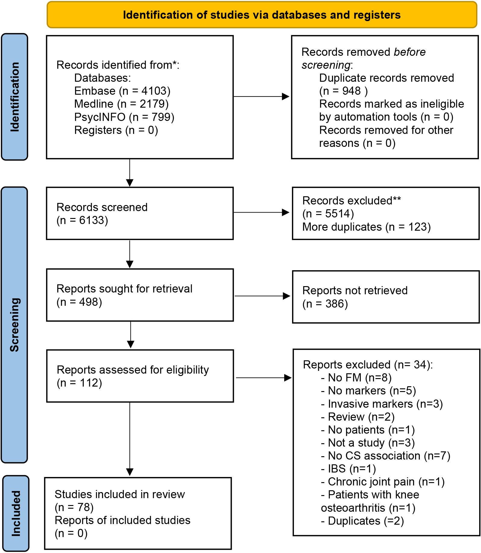
Figure 1.
Flow diagram of study selection process. *Consider, if feasible to do so, reporting the number of records identified from each database or register searched (rather than the total number across all databases/registers). **If automation tools were used, indicate how many records were excluded by a human and how many were excluded by automation tools. From: Page MJ, McKenzie JE, Bossuyt PM, Boutron I, Hoffmann TC, Mulrow CD, et al. The PRISMA 2020 statement: an updated guideline for reporting systematic reviews. BMJ 2021;372:n71. doi: 10.1136/bmj.n71. For more information, visit: http://www.prisma-statement.org/.
The eligibility criteria for the article selection are presented in Table 1. The following inclusion criteria were applied: (1) participants had to be adults (age 18 years or older; (2) patients had to be diagnosed with FM according to the American College of Rheumatology (ACR) criteria of 2010 and with the ACR criteria of 1990 for papers published before 201; (3) HACS markers had to be neurophysiological and non-invasiv; (4) the selected studies were published between 01/01/1994 and 01/04/2022. Articles were excluded if (1) included participants suffered from other forms of pain besides FM (but when patients with FM were compared to patients with other forms of pain besides FM these studies were included); (2) participants suffered from psychiatric comorbidity following specified DSM criteria; (3) the study designs were systematic reviews or meta-analyse; (4) the articles used only invasive markers such as blood tests.
2.3Study selection
The studies were screened (based on title and abstract) by three independent reviewers to exclude studies that were not specific to FM and the study aim. YS screened all, RS screened the first hal; and HT screened the second half. The reviewers subsequently selected articles for inclusion based on full text (
2.4Data extraction
The data extraction process was performed by YS. Two researchers (RS and HT) reviewed the extracted data. The following information was extracted from each study and documented into a table: (1) the study (author names and publication date), (2) the population (number of participants with FM, number of healthy controls (HC) if applicable, age, gender and country, (3) study design, (4) aim of the study, (5) hypotheses, (6) inclusion/exclusion criteria, (7) assessment methods, (8) main findings and (9) definitions of HACS, nociplastic pain or hypersensitivity, when stated in the article.
2.5Risk of bias and quality assessment
Quality assessment of the included articles was carried out using the National Institute of Health (NIH) Quality Assessment Tool for case-control studies, observational cohort and cross-sectional studies, and randomized controlled trials (RCTs) [16]. The NIH tool consists of 13 questions for case-control studies, 14 questions for cross-sectional studies and 14 questions for RCTs. Before assessing all the articles, YS, RS and HT first assessed 6 randomly chosen articles and then discussed it together to determine whether they all deduced the same understanding of the assessment questions. Possible answers for each question of the quality assessment were “ye”, “no”, “cannot determine, not applicable or not reported”. The answer ‘ye’ gave one point, whereas the other answers gave zero points to the study. An overall score between 0 and 13 for case-control studies or 0 and 14 for cross-sectional studies and RCT’s, was then calculated for each included study and the studies were subsequently judged as “good” (score of 75% or above), “fair” (score of 50–75%) or “poor” (score below 50%) quality [17]. Discussions between the three authors were held to solve any encountered disagreements.
The quality of the studies was taken into consideration when interpreting results. Markers identified from studies with a quality of at least ‘fair’ were interpreted as more reliable markers than those identified from studies ranked as ‘poor’ quality. Furthermore, conflicting outcomes from papers studying the same potential marker were considered as inconsistent results, consequently weighing the marker as ‘not valid’.
2.6Study descriptives
The study descriptives of included articles are population (age and sex), country and number of included participants (patients and healthy controls). The results were divided into two main categories based on whether markers were detected by using measurements to assess peripheral or central manifestation of HACS.
3.Results
3.1Search and selection
A total of 78 studies fulfilled the eligibility criteria (Table 1) and were included in this study. Peripheral manifestations of HACS include quantitative sensory testing. Central manifestations of HACS include electrophysiological techniques, conditioned pain modulation, pain anticipation and catastrophizing. Contrary to the peripheral manifestation of HACS, central manifestations are measurements of the CNS, such as brain perfusion using electrophysiological techniques and imaging.
Table 2
Characteristics of included studies (
| Sources (Author; Year) | Population (Gender F/M | Study design | AIM | Hypothesis | Incl./excl. criteria | Altered pain processing or cs or hypersensitivity definition | Assessment method | Results |
|---|---|---|---|---|---|---|---|---|
| Al-Mahdawi et al., 2021 [96] | 31 (23F) patients with FM (18–62 years) and 31 (22F) HC (17–55 years) Iraq | CC | Compare patients with FM and HC with different electrodiagnostic testing and to see whether there is any relationship between the measures | NR | Inclusion: ACR, illness duration from 5 m to 10 y. Exclusion: abnormal upper and lower limb NCSs, EMG and SSR, history of distal symmetrical paresthesia or abnormal sensory examination results, muscle disease, neuromuscular junction disorder, peripheral nerve dysfunction disorders | NR |
|
|
| Baek et al., 2016 [89] | 24(23F) patients with FM (45.21 | CC | To compare cutaneous silent period (CPS) in FM and HC to understand pathophysiology of FM | Inclusion: ACR Exclusion: distal paresthesia, sensory loss, medical condition associated with peripheral neuropathy | NR |
|
|
|
Table 2, continued | ||||||||
|---|---|---|---|---|---|---|---|---|
| Sources (Author; Year) | Population (Gender F/M | Study design | AIM | Hypothesis | Incl./excl. criteria | Altered pain processing or cs or hypersensitivity definition | Assessment method | Results |
| Banic et al., 2004 [57] | 22(18F) patients with FM (mean age 47), 27(19F) whiplash patients (39) and 29(20F) HC (46) Switzerland | CC | To show that Patients with FM and whiplash patients have spinal cord hyperexcitability which causes them to experience severe pain after low intensity nociceptive stimulation | That FM and whiplash patients have facilitated withdrawal reflex | Inclusion: ACR for FM Exclusion: pain for | Decreased reflex threshold indicates spinal cord hypersensitivity |
|
|
| Bendsten et al., 1997 [90] | 25(F) patients with FM (44.9 | CC | To investigate the perception of pain in Patients with FM’ tender muscles | Inclusion: ACR Exclusion: | NR |
|
| |
| Blumenstiel et al., 2011 [34] | 21(F) patients with FM (50.6 | CC | To disclose the similarities and differences in the pathophysiology of FM and CBP | Inclusion: ACR for FM Exclusion: comorbidities (neuropathy, diabetes, infections, disc hernia) | NR |
|
| |
|
Table 2, continued | ||||||||
|---|---|---|---|---|---|---|---|---|
| Sources (Author; Year) | Population (Gender F/M | Study design | AIM | Hypothesis | Incl./excl. criteria | Altered pain processing or cs or hypersensitivity definition | Assessment method | Results |
peripheral sensitization Hand: FM had
| ||||||||
| Bosma et al., 2016 [18] | 20(F) patients with FM (39 | CC | To characterize the fMRI responses in the spinal cord and brainstem that correspond with TSSP in FM compared to HC |
| Inclusion: ACR Exclusion: opioids, NSAIDs | TSSP evoked at lower frequencies |
|
|
|
Table 2, continued | ||||||||
|---|---|---|---|---|---|---|---|---|
| Sources (Author; Year) | Population (Gender F/M | Study design | AIM | Hypothesis | Incl./excl. criteria | Altered pain processing or cs or hypersensitivity definition | Assessment method | Results |
| Bourke et al., 2021 [43] | 19 (16F) patients with FM (36), 19 (13F) patients with CFS (43) and 20 (14F) HC (34) UK | CC | Investigate possible similarity of CS prevalence in patients with CFS and patients with FM compared to HC |
| Inclusion: CFS diagnosis by CDC criteria, chronic and peristent fatigue as primary complaint in CFS, ACR for FM Exclusion: current psychiatric disorders, no comorbid idiopathic pain disorder, somatic syndrome or comorbid disorder of interest in CFS and FM, smoking, BMI | CS is defined by the presence of both enhanced TS and inefficient CPM |
|
|
| Burgmer et al., 2012 [79] | 17(F) patients with FM (52.59 | CC | Differentiate between increased pain ratings and hyperalgesia related to peripheral or |
| Inclusion: ACR Exclusion: psychiatric disorder, other pain origin, pain medication | NR |
|
|
|
Table 2, continued | ||||||||
|---|---|---|---|---|---|---|---|---|
| Sources (Author; Year) | Population (Gender F/M | Study design | AIM | Hypothesis | Incl./excl. criteria | Altered pain processing or cs or hypersensitivity definition | Assessment method | Results |
| central sensitization and to correlate with cerebral activation pattern |
|
|
| |||||
| Burgmer et al., 2009 [71] | 14(F) patients with FM (51 | CC | To investigate whether patients with FM show alterations in brain morphology in areas of the pain matrix vs HC and whether such volumetric changes are consequences of chronic pain | Volumetric changes will be present in brain areas related to medial pain system in FM vs HC | Inclusion: ACR Exclusion: other pain origin, psychiatric disorders | Decreased GMV indicate CS pre-condition |
|
|
|
Table 2, continued | ||||||||
|---|---|---|---|---|---|---|---|---|
| Sources (Author; Year) | Population (Gender F/M | Study design | AIM | Hypothesis | Incl./excl. criteria | Altered pain processing or cs or hypersensitivity definition | Assessment method | Results |
| Chalaye et al., 2012 [29] | 10(F) patients with FM (46.7 | CC | To compare descending pain inhibition, pain sensitivity and ANS reactivity to pain in FM, IBS and HC | IBS and FM share common but graded pathophysiology, both having impaired descending pain inhibition vs HC but greatest in FM. Same for ANS dysfunction | Inclusion: ACR for FM, ROME II for IBS Exclusion: other medical condition, having FM | NR |
|
|
| Cook et al., 2004 [62] | 9(F) patients with FM (37 | CC | To examine the function of nociceptive system in Patients with FM using fMRI |
| Inclusion: ACR and chronic fatigue syndrome in FM Exclusion: pain medication | NR |
|
|
|
Table 2, continued | ||||||||
|---|---|---|---|---|---|---|---|---|
| Sources (Author; Year) | Population (Gender F/M | Study design | AIM | Hypothesis | Incl./excl. criteria | Altered pain processing or cs or hypersensitivity definition | Assessment method | Results |
|
|
| |||||||
| Craggs et al., 2012 [63] | 13(F) patients with FM (43.4 | CC | Examine the effective connectivity among TSSP-related brain regions in FM&HC and compare whether they are connected in a similar manner | Inclusion: ACR Exclusion: abnormal findings, unrelated to FM, analgesic, NSAID, acetaminophen use | Increased influence of brain regions represents CS presence |
|
| |
| De La Coba et al., 2018 [30] | 30(F) patients with FM (52 | CC | To examine whether BP-related pain modulation, indexed by static and dynamic evoked pain responses, is altered in FM vs HC | Inclusion: ACR, HC free of pain Exclusion: CVD, neurological disorders, psychiatric/somatic disease, | NR |
|
| |
|
Table 2, continued | ||||||||
|---|---|---|---|---|---|---|---|---|
| Sources (Author; Year) | Population (Gender F/M | Study design | AIM | Hypothesis | Incl./excl. criteria | Altered pain processing or cs or hypersensitivity definition | Assessment method | Results |
|
| ||||||||
| De La Coba et al., 2017 [31] | 24(F) patients with FM (52.21 | CC | Evaluate degree of pain sensitization elicited by SREP vs pain threshold and tolerance in terms of associations with clinical FM pain ratings (1) and sensitivity and spec in differentiating btw FM and HC (2) | (1) SREP sensitization in FM but not HC (2) FM | Inclusion: ACR, HC free of pain Exclusion: CVD, somatic/psychiatric disease | NR |
|
|
| De Tommaso et al., 2014 [73] | 199 (171F) patients with FM (40.55 | CC | Examine the nociceptive pathways at the peripheral to the central level in FM | Inclusion: ACR Exclusion: | NR |
|
| |
|
Table 2, continued | ||||||||
|---|---|---|---|---|---|---|---|---|
| Sources (Author; Year) | Population (Gender F/M | Study design | AIM | Hypothesis | Incl./excl. criteria | Altered pain processing or cs or hypersensitivity definition | Assessment method | Results |
|
| ||||||||
| Del Paso et al., 2021 [32] | 40 (37F) patients with FM (51.15 | CC | Investigate the cardiac, vasomotor and myocardial branches of the baroreflex function in patients with FM compared to HC | Patients with FM would demonstrate an inverse relationship of BRS and BEI in the 3 branches with clinical pain intensity | Inclusion: ACR Exclusion: cardiovascular, inflammatory, metabolic and neurological diseases, mental disorders | Pain in FM is defined by hypersensitivity of central nociceptive pathways and incomplete pain-inhibiting mechanisms |
|
|
|
Table 2, continued | ||||||||
|---|---|---|---|---|---|---|---|---|
| Sources (Author; Year) | Population (Gender F/M | Study design | AIM | Hypothesis | Incl./excl. criteria | Altered pain processing or cs or hypersensitivity definition | Assessment method | Results |
|
| ||||||||
| Desmeules et al., 2014 [58] | 137 (92.7%F) patients with FM (50.1 | CC | Evaluate whether neurophysiological, psychological and genetic factors are related in FM | CS observed in FM could be associated with COMT polymorphism (which is linked to | Inclusion: ACR, HC free of pain and no CNS disorder Exclusion: analgesics ( | NFR |
|
|
| Desmeules et al., 2003 [53] | 85 (89%F) patients with FM (49 | CC | Determine whether abnormalities of peripheral and central nociceptive sensory input processing exist outside spontaneous pain areas in FM vs HC, by using QST and a neurophysiologic paradigm independent from subjective reports | Inclusion: ACR, HC free of pain Exclusion: specific medical disorders, necessary analgesic use |
|
|
| |
|
Table 2, continued | ||||||||
|---|---|---|---|---|---|---|---|---|
| Sources (Author; Year) | Population (Gender F/M | Study design | AIM | Hypothesis | Incl./excl. criteria | Altered pain processing or cs or hypersensitivity definition | Assessment method | Results |
|
| ||||||||
| Donadel et al., 2021 [81] | 22 (F) patients with FM (47.14 | CC | Compare the cortical activation and deactivation patterns in patients with FM and HC after 2 stimuli through the assessment of HbO and BOLD fNIRS | Peak latency and HbO concentration differences before and after stimuli would be shorter and larger, respectively, in patients with FM than in HC (explaining a faster cortical response in FM) | Inclusion: ACR Exclusion: pregnant participants, history of malignancy or uncompensated chronic disease, history of neuropsychiatric comorbidities, use of certain medication | Central sensitivity syndrome defined as widespread pain and a state of high reactivity amplifying nociceptive stimuli |
|
|
|
Table 2, continued | ||||||||
|---|---|---|---|---|---|---|---|---|
| Sources (Author; Year) | Population (Gender F/M | Study design | AIM | Hypothesis | Incl./excl. criteria | Altered pain processing or cs or hypersensitivity definition | Assessment method | Results |
| Fallon et al., 2013 (Ipsilateral) [60] | 19(F) patients with FM (40.01 | CC | Evaluate cortical activation patterns during mechanical-tactile stimulation in FM and correlate cortical activation changes with clinical symptoms | Patients with FM would report subjective pain and show alterations in | Inclusion: ACR Exclusion: other disorders, CNS medication, analgesics (except paracetamol) | Increased ERD could be a physiological correlate of CS |
|
|
| Fallon et al., 2013 [91] | 16(F) patients with FM (38.5 | CC | Evaluate whether morphological alterations to subcortical brain regions may contribute to pathophysiological mechanisms and pain in FM | Patients with FM would show subcortical abnormalities in shape and volume and that the degree of these changes would correlate with severity of clinical measures (MPTS) | Inclusion: ACR Exclusion: other disorders, analgesics (except paracetamol), CNS medication | NR |
|
|
|
Table 2, continued | ||||||||
|---|---|---|---|---|---|---|---|---|
| Sources (Author; Year) | Population (Gender F/M | Study design | AIM | Hypothesis | Incl./excl. criteria | Altered pain processing or cs or hypersensitivity definition | Assessment method | Results |
|
| ||||||||
| Gentile et al., 2020 [92] | 38(35F) patients with FM (42.1 | CC | To investigate the motor cortical metabolism and changes of LEPs parameters in patients with FM and HC during movement tasks | NR | Inclusion: ACR, right- handed Exclusion: | NR |
|
|
| Gerdle et al., 2010 [93] | 27(F) patients with FM (37 | CC | Investigate differences in neuromuscular control (differential activation | NR | Inclusion: ACR, HC free of pain | NR |
|
|
|
Table 2, continued | ||||||||
|---|---|---|---|---|---|---|---|---|
| Sources (Author; Year) | Population (Gender F/M | Study design | AIM | Hypothesis | Incl./excl. criteria | Altered pain processing or cs or hypersensitivity definition | Assessment method | Results |
| Gerhardt et al., 2016 [41] | 48(24F) CLP patients (59.7 | CC |
| NR | Inclusion for CBP: CBP as main symptom for | NR |
|
|
|
Table 2, continued | ||||||||
|---|---|---|---|---|---|---|---|---|
| Sources (Author; Year) | Population (Gender F/M | Study design | AIM | Hypothesis | Incl./excl. criteria | Altered pain processing or cs or hypersensitivity definition | Assessment method | Results |
| Goubert et al., 2017 [23] | 26(19F) FM (45 | CS | Compare QST assessment in different LBP patient groups with FM and HC, with regard to chronicity | Altered pain processing in severe CLBP but not in RLBP. Mild CLBP in between RLBP and severe CLBP | Inclusion: ACR for FM Exclusion: other specific diseases, antidep or analgesics (except NSAID, paracetamol) | Decreased PPT indicate hypersensitivity |
|
|
| Giesecke et al., 2004 [67] | 16 (12F) FM (45 | To compare sensory testing and fMRI results between idiopa-thic CLBP patients, patients with FM and HC | NR | Inclusion for CLBP: LBP | NR |
|
| |
|
Table 2, continued | ||||||||
|---|---|---|---|---|---|---|---|---|
| Sources (Author; Year) | Population (Gender F/M | Study design | AIM | Hypothesis | Incl./excl. criteria | Altered pain processing or cs or hypersensitivity definition | Assessment method | Results |
in CLBP and FM, but same stimuli caused less pain in HC and
| ||||||||
| Guedj et al., 2007 [70] | 18(F) patients with FM (49 | CC | Investigate brain processing associated with spontaneous pain in FM | Cerebral perfusion abnormalities would show evidence of altered cerebral processing linked to spontaneous pain in FM | Inclusion: ACR Exclusion: psychiatric disease, other medical condition, specific meds | NR |
|
|
| Hazra et al., 2020 [33] | 50 (42F) patients with FM (38.88 | CC (CS?) | Assess and compare central sensitization and autonomic activity in patients with FM and HC | Central nervous system hyper-sensitivity in patients with FM will explain the generalized pain symptoms in FM | Inclusion: ACR Exclusion: psychiatric disorder, regional pain syndromes, hypothyroidism, major systemic infection, condition having an effect on ANS, disorder of cerebral vascular system, connective tissue or peripheral nerve | CS assessed by increase in prefrontal cortical activity by means of fNIRS for oxygenation measures and patient history (VAS, WPI) |
|
|
|
Table 2, continued | ||||||||
|---|---|---|---|---|---|---|---|---|
| Sources (Author; Year) | Population (Gender F/M | Study design | AIM | Hypothesis | Incl./excl. criteria | Altered pain processing or cs or hypersensitivity definition | Assessment method | Results |
|
| ||||||||
| Hurtig et al., 2001 [39] | 29(F) patients with FM (46y) and 21(F) HC (39y) Sweden | CC | Investigate whether Patients with FM can be subgrouped regarding thermal hyperalgesia and if these subgroups differ in clinical appearance | NR | Inclusion: ACR, HC free of pain | NR |
|
|
| Ichesco et al., 2014 [80] | 18(F) patients with FM (35.8 | CC | Investigate whether IC-CC connectivity patterns are seen in FM and whether they are related to the | Differences in IC-CC and IC-IC connectivity would be seen in FM and that it might provide | Inclusion: ACR, | NR |
|
|
|
Table 2, continued | ||||||||
|---|---|---|---|---|---|---|---|---|
| Sources (Author; Year) | Population (Gender F/M | Study design | AIM | Hypothesis | Incl./excl. criteria | Altered pain processing or cs or hypersensitivity definition | Assessment method | Results |
| hyperalgesia in FM | insights into central neural correlates of chronic pain | left AIC and r&l MFgyrus; left PIC and right SFgyrus
| ||||||
| Ichesco et al., 2016 [64] | 12(F) patients with FM (38.5 | CC | Perturb the central pain system with calibrated pressure pain stimuli and monitor changes in fcMRI induced by this acute pain | Patients with FM would display increased fcMRI in regions involved in pain processing after experimental pain vs HC | Inclusion: ACR, | NR |
|
|
| Janal et al., 2016 [21] | 100(F) TMD- only patients (36.3 | CC | Determine whether CS is found preferentially in myofascial TMD patients that have orofacial pain as regional manifestation of FM | NR | Inclusion: ACR Exclusion: controls with face trauma, dental treatment, facial pain | TSSP and pain AS indicate CS |
|
|
|
Table 2, continued | ||||||||
|---|---|---|---|---|---|---|---|---|
| Sources (Author; Year) | Population (Gender F/M | Study design | AIM | Hypothesis | Incl./excl. criteria | Altered pain processing or cs or hypersensitivity definition | Assessment method | Results |
| Jespersen et al., 2007 [35] | 48(F) patients with FM (49y) and 16(F) HC (45y) Denmark | CC | Evaluate the use of cuff pressure algometry (CPA) in FM and to correlate deep-tissue sensitivity assessed by CPA with other FM disease markers | NR | Inclusion: ACR, HC free of pain Exclusion: other rheumatic disease, psychiatric disorder | Decreased PPT indicate hypersensitivity |
|
|
| Kosek et al., 1996 (Sensory) [37] | 10(F) patients with FM (42.7y) and 10(F) HC (42.3y) Sweden | CC | Examine whether sensory abnormalities in FM are generalized or confined to areas with spontaneous pain | If FM pain is due to dysfunction of central processing of somatosensory input (not peripheral) | Inclusion: ACR, normal lab results, HC free of pain | NR |
|
|
|
Table 2, continued | ||||||||
|---|---|---|---|---|---|---|---|---|
| Sources (Author; Year) | Population (Gender F/M | Study design | AIM | Hypothesis | Incl./excl. criteria | Altered pain processing or cs or hypersensitivity definition | Assessment method | Results |
| Kosek et al., 1996 [36] | 14(F) patients with FM (45.6y) and 14(F) HC (36.8y) Sweden | CC | Evaluate influence of submaximal isometric contraction on pressure pain thresholds (PPT) in FM and HC before and after skin hypoesthesia | NR | Inclusion: ACR, normal lab results, HC free of pain | NR |
|
|
| Lee et al., 2018 [59] | 10(F) patients with FM (45.7 | CS | To analyse resting state EEG of Patients with FM to test whether ES is a mechanism involved in the hypersensitivity of FM brains | Explosive synchronization (ES) can be a mechanism of the hypersensitivity in FM brains | Inclusion: ACR, female, 18–65 age range Exclusion: current psychiatric disorder, HADS | ES condition represent brain hypersensitivity |
|
|
|
Table 2, continued | ||||||||
|---|---|---|---|---|---|---|---|---|
| Sources (Author; Year) | Population (Gender F/M | Study design | AIM | Hypothesis | Incl./excl. criteria | Altered pain processing or cs or hypersensitivity definition | Assessment method | Results |
| Lim et al., 2015 [94] | 21(F) patients with FM (49.9 | CC | To investigate intracortical excitability of primary somatosensory cortex (S1) and its potential role in clinical pain in Patients with FM |
| Inclusion: ACR, wides- pread pain | NR |
|
|
| Loggia et al., 2014 [49] | 31(87.1%F) patients with FM (44.0 | CS | To show potential dysregulation in the neural circuitry related to pain experience (anticipation of pain and pain relief) | NR | Inclusion: ACR Exclusion: HC were free of chronic pain, rheumatic disease Exclusion for both: age | NR |
|
|
|
Table 2, continued | ||||||||
|---|---|---|---|---|---|---|---|---|
| Sources (Author; Year) | Population (Gender F/M | Study design | AIM | Hypothesis | Incl./excl. criteria | Altered pain processing or cs or hypersensitivity definition | Assessment method | Results |
| Loggia et al., 2015 [48] | 31(4M) patients with FM (44.0 | CS | To investigate the association between catastrophizing and brain responses to pain anticipation in FM |
| Inclusion: ACR Exclusion: age | NR |
|
|
| Lopez et al., 2014 [77] | 35(F) patients with FM (46.55 | CC | To identify brain response alterations to non-painful sensory stimuli (auditory, visual, tactile) and their association with clinical pain severity |
| Inclusion: ACR, Vision, hearing normal Exclusion HC: neurologic disorders, chronic/acute pain, substance abuse, psych. illness | NR |
|
|
|
Table 2, continued | ||||||||
|---|---|---|---|---|---|---|---|---|
| Sources (Author; Year) | Population (Gender F/M | Study design | AIM | Hypothesis | Incl./excl. criteria | Altered pain processing or cs or hypersensitivity definition | Assessment method | Results |
|
| ||||||||
| Lopez et al., 2017 [61] | 37(F) patients with FM (46.27 6 7.72) and 35(F) HC (43.86 6 6.05) USA | CC | To identify a neurophysiological signature sensitive to FM | NR | Inclusion: normal vision and hearing, ACR Exclusion HC: neurologic disorders, chronic/acute pain, substance abuse, psych. illness history | increased NPSp response indicates enhanced pain processing |
|
|
|
Table 2, continued | ||||||||
|---|---|---|---|---|---|---|---|---|
| Sources (Author; Year) | Population (Gender F/M | Study design | AIM | Hypothesis | Incl./excl. criteria | Altered pain processing or cs or hypersensitivity definition | Assessment method | Results |
| Lorenz et al., 1998 [72] | 10(F) FM and 10 HC(F) (age- matched) Germany | CC |
| NR | NR | NR |
|
|
| Maestu et al., 2013 [76] | 9(F) patients with FM (36.1 | CC | To characterize brain response differences when stimulation pressure is adjusted to subjective levels of pain in both groups | NR | Inclusion FM: ACR, diagnosis | NR |
|
|
| Maestu et al., 2013 (Reduction) [82] | 54(F) patients with FM: 28 simulation group and 26 sham group Age (40.7 | RCT | To test the effect of very low-intensity transcranial magnetic stimulation (TMS) on FM symptoms | NR | Inclusion FM: ACR, diagnosis | NR |
|
|
|
Table 2, continued | ||||||||
|---|---|---|---|---|---|---|---|---|
| Sources (Author; Year) | Population (Gender F/M | Study design | AIM | Hypothesis | Incl./excl. criteria | Altered pain processing or cs or hypersensitivity definition | Assessment method | Results |
|
| ||||||||
| Martinsen et al., 2014 [55] | 29(F) patients with FM (mean age 49.8 years, range 25–64) and 31 HC(F) (mean age 46.3 years, range 20–63) Sweden | CC |
| Already known: SCWT activates dorsal ACC
| Inclusion FM: 20–65 age range, ACR Exclusion: high BP ( | NR |
|
|
| Martucci et al., 2019 [65] | 16 patients with FM (47.13 | CC | To observe altered frequency-depen- dent activity in spinal cord in FM using resting- state fMRI of the cervical spinal cord | Observe signals indicative of increased resting-state activity (hyperactivity) within the cervical spinal cord in FM | Inclusion FM: ACR, symptoms present | NR |
|
|
|
Table 2, continued | ||||||||
|---|---|---|---|---|---|---|---|---|
| Sources (Author; Year) | Population (Gender F/M | Study design | AIM | Hypothesis | Incl./excl. criteria | Altered pain processing or cs or hypersensitivity definition | Assessment method | Results |
|
| sal quadrant C7/C6 in FM
| |||||||
| Matthey et al., 2013 [83] | 77(F) patients with FM: 39 placebo and 38 MLN all doses Switzerland | RCT | To assess the pharmacodynamic activity of miln- acipran (MLN), a serotonin- noradrenaline reuptake inhibitor, at spinal level on Patients with FM by using the NFR procedure and to see whether its properties affect NFR in FM ( | NR | Inclusion: women, | NR |
|
|
|
Table 2, continued | ||||||||
|---|---|---|---|---|---|---|---|---|
| Sources (Author; Year) | Population (Gender F/M | Study design | AIM | Hypothesis | Incl./excl. criteria | Altered pain processing or cs or hypersensitivity definition | Assessment method | Results |
| NFR procedure |
| |||||||
| Mcloughlin et al., 2011 [47] | 16(F) patients with FM and 18(F) HC Age NR USA | CC | To investigate how physical activity affects brain responses to painful stimulation in FM, using fMRI | Hypothesized that self-reported PA and activity measured objectively (accelerometry) would be negatively related to brain activity in areas involved in sensory and affective pain dimensions, and positively related to areas involved in pain modulation | Inclusion HC: no chronic pain Exclusion both: high-dose anti-depressant psychiatric disorders, ACR Exclusion FM: comorbid pain disorder Prior to testing: no exercise for min 48 h, no alcohol for 24 h, no caffeine for 4 h, no smoking for 2 h | NR |
|
|
|
Table 2, continued | ||||||||
|---|---|---|---|---|---|---|---|---|
| Sources (Author; Year) | Population (Gender F/M | Study design | AIM | Hypothesis | Incl./excl. criteria | Altered pain processing or cs or hypersensitivity definition | Assessment method | Results |
| Montoya et al., 2005 [56] | 12(F) patients with FM (50.58 | CC | To analyze pressure pain thresholds (PPT) and event-related potentials (ERP) elicited by emotional words in FM and HC to evaluate the possibility of a cognitive bias in Patients with FM | Hypothesized that Patients with FM would have increased pain sensitivity and enhanced late positive ERP components triggered by pain-related words compared to neutral ones | Inclusion: no medication 24 h before tests (except 4 FM). FM had tender point assessment assessment), ACR Exclusion: | NR |
|
|
| Morris et al., 1998 [54] | 10(F: M ratio 7: 3) patients with FM (56.5 | CC | Show a disturbance of pain modulation in FM by using capsaicin-induced secondary hyperalgesia (CISH) as a marker of abnormal nociceptive processing | NR | Inclusion: ACR Exclusion: drug allergy, eczema or psoriasis | Increased CISH indicates spinal cord hypersensitivity |
|
|
|
Table 2, continued | ||||||||
|---|---|---|---|---|---|---|---|---|
| Sources (Author; Year) | Population (Gender F/M | Study design | AIM | Hypothesis | Incl./excl. criteria | Altered pain processing or cs or hypersensitivity definition | Assessment method | Results |
| Oliva et al., [44] | 20 (18F) patients with FM (mean age 43) and 20 (18F) HC (mean age 35) UK | CC | To analyze whether attentional analgesia was attenuated in patients with FM compared to HC | Patients with FM would show a deficit in attentional analgesia and fMRI would demonstrate where the deficiency originated in the pain modulatory pathway/attentional network | Inclusion: ACR, minimum 6 month diagnosis of FM Exclusion: other chronic painful conditions, pregnancy, history of psychiatric or neurological illness | NR |
|
|
|
Table 2, continued | ||||||||
|---|---|---|---|---|---|---|---|---|
| Sources (Author; Year) | Population (Gender F/M | Study design | AIM | Hypothesis | Incl./excl. criteria | Altered pain processing or cs or hypersensitivity definition | Assessment method | Results |
|
| ||||||||
| Passard et al., 2007 [84] | 30(29F, 1M) patients with FM (52.6 | RCT | To examine the effects of unilateral repetitive transcranial magnetic stimulation (rTMS) of the motor cortex on chronic widespread pain in Patients with FM | Hypothesized that rTMS of motor cortex can diminish chronic widespread pain in Patients with FM | Inclusion: right-handed, | NR |
|
|
| Potvin et al., 2009 [52] | 37 (93%F) patients with FM (50.6 | CC | Investigate the influence of dopamine-related gene polymorphisms on thermal pain thresholds (TPT) and DNIC efficacy in FM and HC | NR | Inclusion: ACR Exclusion: diabetes, lupus, RA, cardiac pathology, substance abuse | NR |
|
|
| Pujol et al., 2009 [78] | 9(F) patients with FM 47.9 | CC | Generate fMRI maps adjusted to brain response duration after assessing brain response to painful pressure in patients with | NR | Inclusion: ACR, no analgesics 72 h prior to fMRI Exclusion: relevant medical or neurological disorder, substance abuse, psychiatric disease | NR |
|
|
|
Table 2, continued | ||||||||
|---|---|---|---|---|---|---|---|---|
| Sources (Author; Year) | Population (Gender F/M | Study design | AIM | Hypothesis | Incl./excl. criteria | Altered pain processing or cs or hypersensitivity definition | Assessment method | Results |
| FM to show activation patterns and correlation with reported pain |
| |||||||
| Price et al., 2002 [25] | 15(F) FM and 14(F) HC 21–65 years age range USA | RCT | First aim: to determine whether cutaneous hyperalgesia of FMS is specific to heat-induced windup of second pain or includes other types of experimental cutaneous pain Second aim: to determine whether the enhanced windup of FMS patients can be modulated by placebo, naloxone, or fentanyl injections |
| Inclusion: | NR |
|
|
|
Table 2, continued | ||||||||
|---|---|---|---|---|---|---|---|---|
| Sources (Author; Year) | Population (Gender F/M | Study design | AIM | Hypothesis | Incl./excl. criteria | Altered pain processing or cs or hypersensitivity definition | Assessment method | Results |
|
| ||||||||
| Schoen et al., 2016 [50] | 16(F) patients with FM (44.9 | CC | To evaluate a novel method to assess CPM in HC and FM subject | NR | Inclusion: ACR Exclusion: medical and psychiatric comorbidities, major depression, schizophrenia, opioid, analgesics | NR |
|
|
| Staud et al., 2008 (Cutaneous) [24] | 14(F) FM (43.4 | CC | To show the role of alterations in central pain sensitization and not peripheral sensitization or rating bias as responsible for TSSP differences between FM and HC | Hypothesized that FM would have pain thres- holds, long dur- ation heat stimuli ratings and repetitive heat pulses ratings similar to HC. But FM would require lower peak T∘ to evoke same TSSP magnitude | Inclusion: ACR Exclusion: analgesic (NSAID included), acetaminophen use | NR | 3 tests: 1. Pain threshold to selective C-fibre stimulation 2. Long duration (30 s) to test contribution of 3 baseline T∘ (BT) (35∘C, 38∘C, and 40∘C) to pain from heat pulse trains 3. TSSP trains of brief (1.5 s), heat pulses at 0.33 Hz adjusting TSSP of FM and HC
|
|
|
Table 2, continued | ||||||||
|---|---|---|---|---|---|---|---|---|
| Sources (Author; Year) | Population (Gender F/M | Study design | AIM | Hypothesis | Incl./excl. criteria | Altered pain processing or cs or hypersensitivity definition | Assessment method | Results |
| Staud et al., 2003 (DNIC) [27] | 11(F) patients with FM (52.9 | CC | Test the effects of DNIC on temporal summation of second pain (previous work has shown that enhanced temporal summation of second pain is a key feature of abnormal central processing in FM | NR | Inclusion: | TSSP indicates abnormal central pain processing |
|
|
| Staud et al., 2015 [85] | 46 patients with FM: 23 patients (21F, 2M) (46.9 | RCT | Use novel QST protocol to characterize effects of milnacipran (which has shown analgesic effectiveness in other clinical trials of FM) on spinal pain pathways, clinical pain and mechanical/heat hyperalgesia in Patients with FM | Hypothesized that milnacipran would reduce clinical pain and mechanical and heat hyperalgesia in FM | Inclusion: | NR |
|
|
|
Table 2, continued | ||||||||
|---|---|---|---|---|---|---|---|---|
| Sources (Author; Year) | Population (Gender F/M | Study design | AIM | Hypothesis | Incl./excl. criteria | Altered pain processing or cs or hypersensitivity definition | Assessment method | Results |
| Staud et al., 2005 [46] | 12(F) patients with FM (48.4 | CC | Determine whether central or peripheral mechanisms are predominantly involved in the abnormal pain modulation in FM | Hypothesized that isometric exercise would reduce experimental muscle and heat pain in HC but would have either no effect or opposite effect in FM | Inclusion: ACR Exclusion: analgesics (NSAID), acetaminophen use | NR |
|
|
|
Table 2, continued | ||||||||
|---|---|---|---|---|---|---|---|---|
| Sources (Author; Year) | Population (Gender F/M | Study design | AIM | Hypothesis | Incl./excl. criteria | Altered pain processing or cs or hypersensitivity definition | Assessment method | Results |
| Staud et al., 2004 [20] | 104 (96F, 8M) patients with FM (47.9 | CC | Show evidence of central sensitization in Patients with FM by maintaining windup (WU) of second pain at lower stimulus frequencies that would not produce WU when delivered alone | Hypothesized that similar to WU, WU-M (maintained) would be enhanced in FM compared to HC | Inclusion: ACR Exclusion: analgesics (NSAID), acetaminophen use | NR |
|
|
| Staud et al., 2003 [26] | 12(F) patients with FM (45.9) and 24(F) HC (40.3) USA | CC | Determine whether temporal summation of deep muscular pain would occur in HC and would be enhanced in FM | NR | Inclusion: ACR Exclusion: analgesics (NSAID), acetaminophen use | NR |
|
|
|
Table 2, continued | ||||||||
|---|---|---|---|---|---|---|---|---|
| Sources (Author; Year) | Population (Gender F/M | Study design | AIM | Hypothesis | Incl./excl. criteria | Altered pain processing or cs or hypersensitivity definition | Assessment method | Results |
| Staud et al., 2007 [19] | 26(F) patients with FM (44.6 | CC | Evaluate the extent of CS in Patients with FM at both ends of the spinal cord by testing TSSP-M and TSSP-aftersensation of heat pain at the upper extremities and lower extremities | Hypothesized that central pain sensitivity would be not only be abnormal but widespread in Patients with FM | Inclusion: ACR Exclusion: analgesics (NSAID also), acetaminophen, narcotic analgesic use | TSSP-M indicates CS |
|
|
| Staud et al., 2008 [66] | 13(F) patients with FM (43.4 | CC | Compare TSSP-related brain responses in Patients with FM and HC | Hypothesized that FM have increased TSSP sensitivity | Inclusion: ACR Exclusion: abnormal findings, analgesic (NSAID included) and acetaminophen use | NR |
|
|
|
Table 2, continued | ||||||||
|---|---|---|---|---|---|---|---|---|
| Sources (Author; Year) | Population (Gender F/M | Study design | AIM | Hypothesis | Incl./excl. criteria | Altered pain processing or cs or hypersensitivity definition | Assessment method | Results |
|
| ||||||||
| Staud et al., 2010 [45] | 34(F) patients with FM (44.6 | CC | Compare the effects of alternating exercise with rest on clinical pain and thermal/mechanical hyperalgesia in FM and HC | Hypothesized repeated periods of strenuous exercise would activate the endogenous pain inhibitory systems in FM and that this would be most evident during short periods of rest | Inclusion: ACR Exclusion: use of analgesics (NSAID included) and acetaminophen | NR |
|
|
| Staud et al., 2012 [40] | 36(35F, 1M) patients with FM, 23(20F, 3M) HC and 24(18F, 6M) LMP USA | CC | To examine how quantitative sensory tests of primary (mechanical) and secondary (thermal) hyperalgesia predict clinical | Hypothesized that measures of mechanical and heat hyperalgesia would reflect relevant factors of peripheral and central pain | Inclusion: | TSSP indicates CS presence |
|
|
|
Table 2, continued | ||||||||
|---|---|---|---|---|---|---|---|---|
| Sources (Author; Year) | Population (Gender F/M | Study design | AIM | Hypothesis | Incl./excl. criteria | Altered pain processing or cs or hypersensitivity definition | Assessment method | Results |
| pain intensity in patients with chronic musculoskeletal pain disorders | processing and clinical pain | amitriptyline |
| |||||
| Staud et al., 2014 [22] | 38(F) patients with FM (49.1 | CC | To better assess individuals’ pain sensitivity by integrating 3 different WU-trains into a single WU-response function (WU-RF) which is representative of central pain sensitivity. And test whether WU, WU-RFs and WU-aftersensations (WU-AS) could predict clinical pain intensity of FM | NR | Inclusion: | Steeper WU-RF slopes indice abnormal central pain sensitivity |
|
|
|
Table 2, continued | ||||||||
|---|---|---|---|---|---|---|---|---|
| Sources (Author; Year) | Population (Gender F/M | Study design | AIM | Hypothesis | Incl./excl. criteria | Altered pain processing or cs or hypersensitivity definition | Assessment method | Results |
| Staud et al., 2021 (Spinal) [87] | 14 (F) patients with FM (37.6 | CC | Analyze spinal cord activation and modulation during TSSP in patients with FM and HC | NR | Inclusion: ACR Exclusion: major medical or neurological illness, major psychiatric disorders and any contraindications for the MRI environment, pregnancy | Enhanced TSSP and pain after sensation |
|
|
| Staud et al., 2021 (Fibro) [98] | 23 (F) patients with FM (46.2 | CC | Analyze whether patients with FM also represent hypersensitivity to sound augmentation | Patients with FM are also hypersensitive to the augmentation of sound and not only to painful stimuli | Inclusion: ACR Exclusion: major medical or neuro- logical illness, psychiatric disease, and any known hearing abnor- malities | NR |
|
|
| Truini et al., 2015 [95] | 20(19F, 1M) patients with FM (aged 27–62) and 15(13F, 2M) HC (aged 25–54) Italy | CC | Compare the excitability in the pain matrices of Patients with FM and HC and to see whether a preceding conditioning C-fibre LEP reduced the | NR | Inclusion: | NR |
|
|
|
Table 2, continued | ||||||||
|---|---|---|---|---|---|---|---|---|
| Sources (Author; Year) | Population (Gender F/M | Study design | AIM | Hypothesis | Incl./excl. criteria | Altered pain processing or cs or hypersensitivity definition | Assessment method | Results |
| following A |
| |||||||
| Van Vliet et al., 2018 [38] | 34(24F, 10M) DM2 patients (54 | CC | To assess pain prevalence, severity and characteristics in patients with myotonic dystrophy type 2 (DM2) and compare them with FM and HC subjects | NR | Inclusion: ACR Exclusion: | CS is less prominent in DM2 patients vs FM, which confirms the presence of CS in FM |
|
|
| Vaegter et al., 2016 [51] | 400(263F, 137M) chronic pain patients (48 | CC | To see if there are different subgroups in a cohort of patients with different chronic pain conditions and to investigate differences in pain and pain hypersensitivity between these subgroups | NR | Inclusion: | NR |
|
|
|
Table 2, continued | ||||||||
|---|---|---|---|---|---|---|---|---|
| Sources (Author; Year) | Population (Gender F/M | Study design | AIM | Hypothesis | Incl./excl. criteria | Altered pain processing or cs or hypersensitivity definition | Assessment method | Results |
|
| ||||||||
| Van Assche et al., 2020 [74] | 92(63F, 29M) patients with FM (49 | C | To estimate the prevalence of thermo-nociceptive system dysfunction using LEPs in Patients with FM | Hypothesized that small fibre neuropathy (SFN) is a significant contributor to the pathophysiology of FM (not supported by results) | Inclusion: ACR Exclusion: | NR |
|
|
| Vecchio et al., 2020 [75] | 81 (73F) patients with FM (50 | C | Analyze the functional changes of central nociceptive pathways measured by LEP’s and the correlation with clinical characteristics | NR | Inclusion: ACR, age between 18–75 years Exclusion: education below 8 years and any cause of PNS or CNS diseases, psychiatric conditions other than anxiety and depression disorders according to the DSM V, active malignancies or history of cancer, use of drugs acting on the CNS and chronic opioid therapy | NR |
|
|
| Wik et al., 2006 [69] | 8(F) patients with FM (42–56 years) Sweden | C | Analysis of PET scan measure of regional cerebral blood flow (rCBF) during externally induced acute pain and rest in patients with FM | NR | Inclusion: ACR | NR |
|
|
|
Table 2, continued | ||||||||
|---|---|---|---|---|---|---|---|---|
| Sources (Author; Year) | Population (Gender F/M | Study design | AIM | Hypothesis | Incl./excl. criteria | Altered pain processing or cs or hypersensitivity definition | Assessment method | Results |
| Wik et al., 2003 [68] | 8(F) patients with FM (42–56 years) and 8(F) HC (27–42 years) Norway | CS | To study the CNS in FM and compare PET scan measures of rCBF in FM and HC subjects at rest | NR | Inclusion: ACR Exclusion: organic brain disorder, somatic disease | NR |
|
|
| Wodehouse et al., 2018 [88] | 14(13F, 1M) patients with FM (46.7 | CS | To see whether QST detects changes in pain thresholds of Patients with FM receiving pregabalin treatment | NR | Inclusion: | NR |
|
|
|
Table 2, continued | ||||||||
|---|---|---|---|---|---|---|---|---|
| Sources (Author; Year) | Population (Gender F/M | Study design | AIM | Hypothesis | Incl./excl. criteria | Altered pain processing or cs or hypersensitivity definition | Assessment method | Results |
| Zhang et al., 2015 [86] | 121(63F, 58M) chronic pain pt on opioid therapy (46.6 | CS | To compare the sensitivity to experimental pain of chronic pain patients on opioid therapy vs chronic pain patients non non-opioid therapy and HC by using QST | NR | Inclusion: pain free HC and no opioid treatment min 6 m, pain non-opioid: stable pain condition but no opioid treatment for min 3 m, pain opioid: stable pain condition for min 3m Exclusion: sensory deficits at a QST site, interventions altering QST response, psychiatric illness | NR |
|
|
ACR: American college of rheumatology FM diagnosis criteria, AIC: anterior IC, ACC: anterior cingulate cortex, aMCC: anterior mid-cingulate cortex, APA: action potential amplitude, AUC: area under the curve, AS: pain after-sensation, ANS: autonomic nervous system, BP: blood-pressure, BOLD: blood-oxygen-level-dependent, BDI: Beck depression inventory, BP-PCS: Brazilian Portuguese Profile of chronic pain: screen, BPI: brief pain inventory, BRS; baroreflex sensitivity, BEI: baroreflex effectiveness, CC: cingulate cortex, C: cohort, cor: correlation, CLBP: chronic low back pain, CPM: conditioned pain modulation, CC: case control, CS: cross-sectional, CLP: chronic localized pain, CWP: chronic widespread pain, CSP: cutaneous silent period, CNS: central nervous system, CBP: chronic back pain, CLBP: chronic low back pain, CS: central-sensitization, C(D)T: cold (detection) threshold, CES-D: Center for Epidemiological Studies Depression Scale, CPT: cold pain threshold, CDC; centers for disease control, CFQ: Chalder fatigue questionnaire, CFS: chronic fatigue syndrome, CSS: central sensitization symptoms, CSI: central sensitization inventory, DM2: myotonic dystrophy type 2, DNIC: diffuse noxious inhibitory control, dif: difference, DLPFC: dorsolateral prefrontal, DEPS: depression scale, DBP: diastolic blood pressure, DBT: deep breathing test, DSM V: diagnostic and statistical manual of mental disorders, ES: explosive synchronization, EQ-5 L-5D: EuroQol The 5-level EQ-5D version, ED: electrodiagnostic, EDA: electrodermal activity, EPTT: electric pain tolerance threshold, EMG: electromyography, freq: frequency, FM: patients with fibromyalgia, fNIRS: functional near-infrared spectroscopy, FSS: fatigue severity scale, FP: frontopolar cortex, GMV: gray matter volume, HADS: hospital anxiety and depression scale, HbO: oxyhemoglobin, HC: healthy controls, HPT, heat pain threshold, HR: heart rate, HRV: heart rate variability, IBI: interbeat interval, ISI: interstimulus interval, IPL: inferior parietal lobule, IC: insular cortex, IPAQ: international physical activity questionnaire, IBS: irritable bowel syndrome, ILC: ipsilateral locus coeruleus, PDI: pain disability index, vs: compared to, pt: patients, y: years, w: weeks, VAS: visual analogue scale, LMP: local musculoskeletal pain, LOC: lateral occipital cortex, lPFC: lateral prefrontal cortex, MC: motor cortex, MPT: mechanical pain threshold, MPS: mechanical pain sensitivity, MVC: maximum voluntary contraction, MCV: medical college of Virginia pain questionnaire, MDT: mechanical detection threshold, MPQ: McGill pain questionnaire, MPFC: medial prefrontal cortex, MRS: multiple random staircase method, M1: primary motor cortex, NFR: nociceptive flexion reflex, NRS: numerical rating scale, NPQ: neuropathic pain questionnaire, OQS: Oviedo quality of sleep questionnaire, OFC: orbitofrontal cortices, stim: stimulation, OP: occipital pole, SF-MPQ: short-form of the McGill pain questionnaire, periph: peripheral, P-Ins: posterior insula, PPT: pressure pain threshold, PPI: present pain intensity, PPC: posterior parietal cortex, PS: peripheral sensitization, PAG; periaqueductal grey, PEP: pre-ejection period, PSQ-3: Pain and Sleep Questionnaire Three-Item, PASS: Pain anxiety symptom scales, PCS: pain catastrophizing scale, PrCG: pre-central gyrus, PCC: posterior cingulate cortex, Precun: precuneus, PL: paracentral lobule, PFC: pre-frontal cortex, QoL: quality of life, rCBF: regional cerebral blood flow, RLBP: recurrent low back pain, RCT: randomized controlled trial, ROI: region of interest, RSVP: rapid serial visual presentation, RVM: rostral ventromedial medulla, RMDQ: Roland-Morris Disability Questionnaire, ROC: receiver operator characteristics, SSR: sympathetic skin response, SREP: slowly repeated evoked pain, SPECT: single-photon emission computed tomography, signif: significant, S1&S2: primary and secondary somatosensory cortices, SMC: sensorimotor cortex, STPI: State-Trait personality Inventory (to assess anxiety), SF-36-PF: physical function subscale of the SF-36, SBP: systolic blood pressure, SPL: superior parietal lobule, STAI: State-trait anxiety inventory, SV: stroke volume, TS: temporal summation, , TSSP: temporal summation of second pain, T-T: threshold/tolerance, thal: thalamus, TMD: temporo-mandibular disorder, TPT: thermal pain threshold, cortex, TPR: total peripheral resistance, TSK: Tampa scale of kinesiophobia, VBM: voxel-based morphometry, VDT: vibration detection threshold, VGEE: generalized estimating equations, W(D)T: warm (detection) threshold, WU: wind up of pain, WU-AS: wind-up pain after-sensation, WPI: widespread pain index, +: positive, -: negative, r & l: right & left, ipsilat/contralat: ipsilateral/contralateral,
3.2Study characteristics
The study characteristics are shown in Table 2. In total, 2383 patients with FM, 1766 Healthy Controls (HC), and 1085 patients with other chronic pain conditions were included.
Peripheral manifestations of HACS were shown in the following studies: temporal summation of secondary pain (TSSP) and pain after-sensations (AS) were studied in eleven studies [18, 19, 20, 21, 22, 23, 24, 25, 26, 27, 28], the autonomic nervous system and slowly repeated evoked pain (SREP) sensitization were studied in five studies [29, 30, 31, 32, 33], quantitative sensory testing (QST) measures (heat, pressure and mechanical and sound ‘pressure’ pain thresholds) were used in thirteen studies [21, 23, 34, 35, 36, 37, 38, 39, 40, 41, 42, 43, 44] and the motor activity and FM was studied in four studies [36, 45, 46, 47].
Central manifestations of HACS were shown in the following studies: pain anticipation was studied twice [48, 49], conditioned pain modulation (CPM) was studied nine times [27, 29, 38, 43, 50, 51, 52, 53, 54], and three studies reported on the effect of distraction on pain [44, 55, 56], electrophysiological techniques were used in twenty-two studies [28, 44, 48, 49, 53, 55, 56, 57, 58, 59, 60, 61, 62, 63, 64, 65, 66, 67, 68, 69, 70, 71], laser-evoked potential (LEP) amplitudes were applied in four studies [72, 73, 74, 75], and brain region activation to stimuli and brain region connectivity were studied in sixteen studies [28, 33, 44, 60, 61, 62, 63, 64, 66, 69, 76, 77, 78, 79, 80, 81].
Table 3
Risk of bias assessment of included studies (
|
Table 3a. Case control studies ( | ||||||||||||||||
|---|---|---|---|---|---|---|---|---|---|---|---|---|---|---|---|---|
| Study | Q1 | Q2 | Q3 | Q4 | Q5 | Q6 | Q7 | Q8 | Q9 | Q10 | Q11 | Q12 | Q13 | Score | % | Quality |
| Al-Mahdawi et al. [96] | Yes | Yes | No | No | CD | Yes | No | NR | NA | Yes | CD | NR | No | 4/13 | 31% | Poor |
| Baek et al. [89] | Yes | No | NR | No | NR | NR | No | NA | NR | Yes | No | NR | No | 2/13 | 15% | Poor |
| Banic et al. [57] | Yes | No | NR | Yes | NR | No | No | NA | NR | Yes | Yes | NR | Yes | 5/13 | 38% | Poor |
| Bendsten et al. [90] | Yes | No | NR | No | NR | Yes | Yes | NA | NR | Yes | Yes | NR | No | 5/13 | 38% | Poor |
| Blumenstiel et al. [34] | Yes | No | NR | No | No | No | No | No | NR | Yes | Yes | NR | Yes | 4/13 | 31% | Poor |
| Bosma et al. [18] | Yes | No | NR | No | No | No | Yes | NA | NR | Yes | Yes | NR | No | 4/13 | 31% | Poor |
| Bourke et al. [43] | Yes | No | No | No | No | NR | Yes | NR | NA | Yes | Yes | NR | No | 4/13 | 31% | Poor |
| Burgmer et al. [79] | Yes | No | NR | No | No | Yes | Yes | NA | NR | Yes | Yes | NR | Yes | 6/13 | 46% | Poor |
| Burgmer et al. [71] | Yes | No | NR | No | NR | No | No | NA | NR | Yes | Yes | NR | Yes | 4/13 | 31% | Poor |
| Chalaye et al. [29] | Yes | No | NR | No | NR | Yes | Yes | NA | NR | Yes | Yes | NR | No | 5/13 | 38% | Poor |
| Cook et al. [62] | Yes | No | NR | No | Yes | Yes | Yes | NA | NR | Yes | Yes | NR | No | 6/13 | 46% | Poor |
| Craggs et al. [63] | Yes | No | NR | No | No | No | No | NA | NR | Yes | Yes | NR | No | 3/13 | 23% | Poor |
| De la Coba et al. [30] | Yes | No | NR | No | Yes | Yes | Yes | NA | NR | Yes | Yes | Yes | Yes | 8/13 | 61% | Fair |
| De la Coba et al. [31] | Yes | No | NR | No | Yes | Yes | Yes | NA | NR | Yes | Yes | NR | Yes | 7/13 | 54% | Fair |
| De Tommaso et al. [73] | Yes | Yes | NR | No | Yes | Yes | No | NA | NR | Yes | Yes | Yes | Yes | 8/13 | 61% | Fair |
| Desmeules et al. [58] | Yes | No | NR | No | Yes | Yes | Yes | NA | NR | Yes | Yes | NR | No | 6/13 | 46% | Poor |
| Desmeules et al. [53] | Yes | No | NR | No | Yes | Yes | Yes | NA | NR | Yes | Yes | NR | No | 6/13 | 46% | Poor |
| Del Paso et al. [32] | No | Yes | No | No | NR | Yes | Yes | NR | NA | Yes | Yes | NR | No | 5/13 | 38% | Poor |
| Donadel et al. [81] | Yes | Yes | Yes | Yes | CD | Yes | Yes | NR | NA | Yes | Yes | NR | Yes | 9/13 | 69% | Fair |
| Fallon et al. [60] | Yes | No | NR | No | No | Yes | Yes | NA | NR | Yes | Yes | NR | No | 5/13 | 38% | Poor |
| Fallon et al. [91] | Yes | No | NR | No | No | Yes | Yes | NA | NR | Yes | Yes | NR | Yes | 6/13 | 46% | Poor |
| Gentile et al. [92] | Yes | No | NR | No | Yes | Yes | Yes | NA | NR | Yes | Yes | NR | No | 6/13 | 46% | Poor |
| Gerdle et al. [93] | Yes | No | NR | No | No | Yes | Yes | NA | NR | Yes | Yes | NR | No | 5/13 | 38% | Poor |
| Gerhardt et al. [41] | Yes | No | NR | No | NR | Yes | Yes | NA | NR | Yes | Yes | Yes | Yes | 7/13 | 54% | Fair |
| Giesecke et al. [67] | No | No | NR | No | No | Yes | Yes | NA | NR | Yes | Yes | NR | No | 4/13 | 30% | Poor |
| Goubert et al. [23] | Yes | No | NR | No | Yes | Yes | Yes | NA | NR | Yes | Yes | NR | Yes | 7/13 | 54% | Fair |
| Guedj et al. [70] | Yes | No | NR | No | No | No | No | NA | NR | Yes | No | NR | No | 2/13 | 15% | Poor |
| Hazra et al. [33] | Yes | No | Yes | NR | NR | Yes | NR | NR | NA | Yes | Yes | NR | No | 5/13 | 38% | Poor |
| Hurtig et al. [39] | Yes | No | NR | No | Yes | No | Yes | NA | NR | Yes | Yes | NR | No | 5/13 | 38% | Poor |
| Ichesco et al. [80] | Yes | No | NR | No | Yes | Yes | Yes | NA | NR | Yes | Yes | NR | No | 6/13 | 46% | Poor |
| Ichesco et al. [64] | Yes | No | NR | No | NR | Yes | Yes | NA | NR | Yes | Yes | NR | No | 5/13 | 38% | Poor |
| Janal et al. [21] | Yes | Yes | NR | Yes | Yes | Yes | Yes | NA | NR | Yes | Yes | No | No | 8/13 | 61% | Fair |
| Jespersen et al. [35] | Yes | No | NR | No | Yes | Yes | Yes | NA | NR | Yes | Yes | NR | No | 6/13 | 46% | Poor |
| Kosek et al. [37] | Yes | No | NR | No | No | No | No | NA | NR | Yes | Yes | NR | No | 3/13 | 23% | Poor |
| Kosek et al. [36] | Yes | No | NR | No | No | No | No | NA | NR | Yes | Yes | NR | No | 3/13 | 23% | Poor |
| Lim et al. [94] | Yes | No | NR | No | No | Yes | Yes | NA | NR | Yes | Yes | NR | Yes | 6/13 | 46% | Poor |
| Loggia et al. [49] | Yes | Yes | NR | No | No | Yes | Yes | NA | NR | Yes | Yes | NR | Yes | 7/13 | 54% | Fair |
| Lopez et al. [77] | Yes | No | NR | No | Yes | Yes | Yes | NA | NR | Yes | Yes | NR | No | 6/13 | 46% | Poor |
| Lopez et al. [61] | Yes | Yes | NR | No | Yes | Yes | Yes | NA | NR | Yes | Yes | NR | Yes | 8/13 | 61% | Fair |
| Lorenz et al. [72] | Yes | No | NR | No | No | CD | No | NA | NR | CD | No | NR | No | 1/13 | 7% | Poor |
| Maestu et al. [76] | Yes | No | NR | No | CD | Yes | Yes | NA | NR | Yes | Yes | NR | No | 5/13 | 38% | Poor |
| Martinsen et al. [55] | Yes | No | NR | No | No | No | No | NA | NR | Yes | Yes | NR | No | 3/13 | 23% | Poor |
| Martucci et al. [65] | Yes | No | NR | No | CD | Yes | Yes | NA | NR | Yes | Yes | NR | No | 5/13 | 38% | Poor |
|
Table 3a, continued | ||||||||||||||||
|---|---|---|---|---|---|---|---|---|---|---|---|---|---|---|---|---|
| Study | Q1 | Q2 | Q3 | Q4 | Q5 | Q6 | Q7 | Q8 | Q9 | Q10 | Q11 | Q12 | Q13 | Score | % | Quality |
| Mcloughlin et al. [47] | Yes | No | NR | No | No | Yes | Yes | NA | NR | Yes | Yes | NR | Yes | 6/13 | 46% | Poor |
| Montoya et al. [56] | Yes | No | NR | No | No | Yes | Yes | NA | NR | Yes | Yes | NR | No | 5/13 | 38% | Poor |
| Morris et al. [54] | Yes | No | NR | No | No | Yes | Yes | NA | NR | Yes | Yes | NR | No | 5/13 | 38% | Poor |
| Oliva et al. [44] | Yes | No | No | NR | No | Yes | Yes | NA | No | Yes | Yes | NR | Yes | 6/13 | 46% | Poor |
| Potvin et al. [52] | Yes | No | NR | No | No | No | No | NA | NR | Yes | Yes | NR | Yes | 4/13 | 31% | Poor |
| Price et al. [25] | Yes | No | NR | No | NR | Yes | Yes | NA | NR | Yes | Yes | Yes | No | 6/13 | 46% | Poor |
| Pujol et al. [78] | Yes | No | NR | No | Yes | Yes | Yes | NA | NR | Yes | Yes | NR | Yes | 7/13 | 54% | Fair |
| Schoen et al. [50] | Yes | No | NR | No | No | Yes | Yes | NA | NR | Yes | Yes | NR | No | 5/13 | 38% | Poor |
| Staud et al. [24] | Yes | No | NR | No | No | Yes | Yes | NA | NR | Yes | Yes | NR | No | 5/13 | 38% | Poor |
| Staud et al. [27] | Yes | No | NR | No | Yes | Yes | Yes | NA | NR | Yes | Yes | NR | Yes | 7/13 | 54% | Fair |
| Staud et al. [46] | Yes | No | NR | No | No | Yes | No | NA | NR | Yes | Yes | NR | No | 4/13 | 31% | Poor |
| Staud et al. [20] | Yes | No | NR | No | No | No | No | NA | NR | Yes | Yes | NR | No | 3/13 | 23% | Poor |
| Staud et al. [26] | Yes | No | NR | No | No | No | No | NA | NR | Yes | Yes | NR | No | 3/13 | 23% | Poor |
| Staud et al. [19] | Yes | No | NR | No | No | No | No | NA | NR | Yes | Yes | NR | No | 3/13 | 23% | Poor |
| Staud et al. [66] | Yes | No | NR | No | No | No | No | NA | NR | Yes | Yes | NR | No | 3/13 | 23% | Poor |
| Staud et al. [45] | Yes | No | NR | No | CD | No | No | NA | NR | Yes | Yes | NR | No | 3/13 | 23% | Poor |
| Staud et al. [40] | Yes | No | NR | No | No | Yes | Yes | NA | NR | Yes | Yes | NR | Yes | 6/13 | 46% | Poor |
| Staud et al. [22] | Yes | No | NR | Yes | CD | Yes | Yes | NA | NR | Yes | Yes | NR | No | 6/13 | 46% | Poor |
| Staud et al. [28] | Yes | No | NR | NR | Yes | Yes | Yes | NA | No | Yes | Yes | NR | Yes | 7/13 | 54% | Fair |
| Staud et al. [42] | Yes | No | CD | NR | Yes | Yes | Yes | NA | No | Yes | Yes | NR | Yes | 7/13 | 54% | Fair |
| Truini et al. [95] | Yes | No | NR | No | CD | Yes | Yes | NA | NR | Yes | Yes | NR | No | 5/13 | 38% | Poor |
| Van Vliet et al. [38] | Yes | No | NR | No | No | Yes | Yes | NA | NR | Yes | Yes | NR | Yes | 6/13 | 46% | Poor |
Q: question, NR: not reported, NA: not applicable, CD: cannot determine. The quality of included studies was assessed using the National Institute of Health (NIH) Quality Assessment Tool for Case Control Studies (https: //www.nhlbi.nih.gov/health-topics/study-quality-assessment-tools). Q1: Was the research question or objective in this paper clearly stated and appropriate? Q2: Was the study population clearly specified and defined? Q3: Target population and case representation, Q4: Did the authors include a sample size justification? Q5: Were controls selected or recruited from the same or similar population that gave rise to the cases (including the same timeframe)? Q6: Were the definitions, inclusion and exclusion criteria, algorithms or processes used to identify or select cases and controls valid, reliable, and implemented consistently across all study participants? Q7: Were the cases clearly defined and differentiated from controls? Q8: If less than 100 percent of eligible cases and/or controls were selected for the study, were the cases and/or controls randomly selected from those eligible? Q9: Was there use of concurrent controls? Q10: Were the investigators able to confirm that the exposure/risk occurred prior to the development of the condition or event that defined a participant as a case? Q11: Were the measures of exposure/risk clearly defined, valid, reliable, and implemented consistently (including the same time period) across all study participants? Q12: Were the assessors of exposure/risk blinded to the case or control status of participants? Q13: Were key potential confounding variables measured and adjusted statistically in the analyses? If matching was used, did the investigators account for matching during study analysis?
|
Table 3b. Cross-sectional studies ( | |||||||||||||||||
|---|---|---|---|---|---|---|---|---|---|---|---|---|---|---|---|---|---|
| Study | Q1 | Q2 | Q3 | Q4 | Q5 | Q6 | Q7 | Q8 | Q9 | Q10 | Q11 | Q12 | Q13 | Q14 | Score | % | Quality |
| Lee et al. [59] | Yes | No | Yes | Yes | No | No | No | Yes | Yes | No | Yes | NR | NA | No | 6/14 | 43% | Poor |
| Loggia et al. [48] | Yes | Yes | Yes | No | No | No | No | No | Yes | No | Yes | NR | NA | Yes | 6/14 | 43% | Poor |
| Vaegter et al. [51] | Yes | Yes | NR | Yes | NR | No | No | NA | Yes | Yes | Yes | NR | NA | Yes | 7/14 | 50% | Fair |
| Van Assche et al. [74] | Yes | Yes | No | Yes | No | No | No | No | Yes | No | Yes | NR | NA | Yes | 6/14 | 43% | Poor |
| Vecchio et al. [75] | No | Yes | Yes | NR | CD | Yes | No | Yes | No | Yes | NR | Yes | Yes | Yes | 8/14 | 57% | Fair |
| Wik et al. [69] | Yes | No | NR | Yes | No | No | No | No | Yes | No | Yes | NR | NA | No | 4/14 | 29% | Poor |
| Wik et al. [68] | Yes | No | NR | Yes | No | No | No | NA | Yes | No | Yes | NR | NA | No | 4/14 | 29% | Poor |
| Wodehouse et al. [88] | Yes | No | Yes | CD | No | No | No | No | Yes | Yes | Yes | NR | NA | No | 5/14 | 36% | Poor |
| Zhang et al. [86] | Yes | No | Yes | Yes | No | No | No | Yes | Yes | Yes | Yes | Yes | NA | No | 8/14 | 57% | Fair |
Q: question, NR: not reported, NA: not applicable. The quality of included studies was assessed using the National Institute of Health (NIH) Quality Assessment Tool for Observational Cohort and Cross-Sectional Studies (https: //www.nhlbi.nih.gov/health-topics/study-quality-assessment-tools). Q1: Was the research question or objective in this paper clearly stated? Q2: Was the study population clearly specified and defined? Q3: Was the participation rate of eligible persons at least 50%? Q4: Were all the subjects selected or recruited from the same or similar populations (including the same time period)? Were inclusion and exclusion criteria for being in the study prespecified and applied uniformly to all participants? Q5: Was a sample size justification, power description, or variance and effect estimates provided? Q6: For the analyses in this paper, were the exposure(s) of interest measured prior to the outcome(s) being measured? Q7: Was the timeframe sufficient so that one could reasonably expect to see an association between exposure and outcome if it existed? Q8: For exposures that can vary in amount or level, did the study examine different levels of the exposure as related to the outcome (e.g., categories of exposure, or exposure measured as continuous variable)? Q9: Were the exposure measures (independent variables) clearly defined, valid, reliable, and implemented consistently across all study participants? Q10: Was the exposure(s) assessed more than once over time? Q11: Were the outcome measures (dependent variables) clearly defined, valid, reliable, and implemented consistently across all study participants? Q12: Were the outcome assessors blinded to the exposure status of participants? Q13: Was loss to follow-up after baseline 20% or less? Q14: Were key potential confounding variables measured and adjusted statistically for their impact on the relationship between exposure(s) and outcome(s)?
|
Table 3c. RCT studies ( | |||||||||||||||||
|---|---|---|---|---|---|---|---|---|---|---|---|---|---|---|---|---|---|
| Study | Q1 | Q2 | Q3 | Q4 | Q5 | Q6 | Q7 | Q8 | Q9 | Q10 | Q11 | Q12 | Q13 | Q14 | Score | % | Quality |
| Maestu et al. [82] | Yes | Yes | Yes | Yes | Yes | Yes | Yes | Yes | Yes | Yes | Yes | No | Yes | Yes | 13/14 | 93% | Good |
| Matthey et al. [83] | Yes | Yes | Yes | Yes | Yes | Yes | Yes | Yes | Yes | Yes | Yes | Yes | No | Yes | 13/14 | 93% | Good |
| Passard et al. [84] | Yes | Yes | Yes | Yes | Yes | Yes | Yes | Yes | Yes | Yes | Yes | No | Yes | Yes | 13/14 | 93% | Good |
| Staud et al. [85] | Yes | Yes | Yes | Yes | Yes | Yes | No | Yes | Yes | Yes | Yes | Yes | Yes | Yes | 13/14 | 93% | Good |
The quality of included studies was assessed using the National Institute of Health (NIH) Quality Assessment Tool for Controlled Intervention Studies (https: //www.nhlbi.nih.gov/health-topics/study-quality-assessment-tools). Q1: Was the study described as randomized, a randomized trial, a randomized clinical trial, or an RCT? Q2: Was the method of randomization adequate (i.e., use of randomly generated assignment)? Q3: Was the treatment allocation concealed (so that assignments could not be predicted)? Q4: Were study participants and providers blinded to treatment group assignment? Q5: Were the people assessing the outcomes blinded to the participants’ group assignments? Q6: Were the groups similar at baseline on important characteristics that could affect outcomes (e.g., demographics, risk factors, comorbid conditions)? Q7 Was the overall drop-out rate from the study at endpoint 20% or lower of the number allocated to treatment? Q8: Was the differential drop-out rate (between treatment groups) at endpoint 15 percentage points or lower? Q9: Was there high adherence to the intervention protocols for each treatment group? Q10: Were other interventions avoided or similar in the groups (e.g., similar background treatments)? Q11: Were outcomes assessed using valid and reliable measures, implemented consistently across all study participants? Q12: Did the authors report that the sample size was sufficiently large to be able to detect a difference in the main outcome between groups with at least 80% power? Q13: Were outcomes reported or subgroups analyzed prespecified (i.e., identified before analyses were conducted)? Q14: Were all randomized participants analyzed in the group to which they were originally assigned, i.e., did they use an intention-to-treat analysis?
3.3Risk of bias and quality assessment
The average risk of bias assessment scores was 39% for case control studies (
3.4Measurements to assess peripheral manifestations of HACS
3.4.1Quantitative sensory testing (QST)
Table 4
Summary of temporal summation of second pain, pain after-sensation and pressure pain threshold findings
| TSSP | Stimulus frequency eliciting TSSP | After sensations | Rate of WU after sensation decline | Pressure pain threshold | |
|---|---|---|---|---|---|
| Patients with FM |
| ||||
| Healthy controls |
|
|
|
|
|
Table 5
Summary of electrophysiological technique findings in patients with fibromyalgia
| EMG | EEG | fMRI | PET | VBM | SPECT | fNIRS |
|---|---|---|---|---|---|---|
| Explosive synchronization conditions in resting state EEG | Hypoperfusion: bilateral frontal, ant/post cingulate, med temporal cerebellar cortices Hyperperfusion: S1 & S2 |
FM: fibromyalgia,
Temporal summation of second pain (TSSP) and after-sensations (AS): TSSP, also known as windup (WU), is a process evoked by repetitive harmless stimuli supposed to cause higher excitability of the dorsal horn neurons, mediated by C nociceptive fibers. Pressure algometry was used in three studies [23, 26, 43] and thermodes were used in seven studies [18, 19, 20, 21, 25, 27, 28] for pressure and heat pain stimulation, respectively, to measure TSSP. Higher TSSP ratings [19], no difference in TSSP ratings [28], higher pain AS [18, 26], slower rate of AS decline [19, 20, 21] and lower stimulus temperature and frequency needed to elicit TSSP in patients [18, 24, 66] were demonstrated and are, according to our division, signs associated with HACS. The authors of eight studies showed higher TSSP sensitivity in patients with FM [19, 20, 23, 25, 26, 27, 28, 43] compared to HC. No TSSP difference between groups was reported in two studies [18, 21]. Results are shown in Table 4.
Pain thresholds: Statistically significant lower pressure pain thresholds (PPT) [23, 34, 35, 36, 37, 41, 42, 43, 44], heat pain thresholds (HPT) [34, 41, 42, 43, 44], cold pain thresholds (CPT) [34, 37, 39, 43, 44], mechanical pain thresholds (MPT) [34] and electrical pain thresholds (EPT) [38] and sound ‘pressure’ threshold [42] in patients with FM compared to HC were demonstrated. Janal and co-workers observed no statistically significant differences in thermal pain thresholds between patients with temporo-mandibular joint disorder with FM and patients with temporo-mandibular joint disorder without FM [21]. Patients with FM displayed higher warmth detection thresholds (WDT) compared to HC [44]. Bourke and colleagues however showed no difference for WDT and cold detection threshold (CDT) between groups [43].
3.4.2Other peripheral measurements
Other markers of HACS include slowly repeated evoked pain (SREP) sensitization, the autonomic nervous system (ANS) response to pain measured with an electrocardiogram, electromyography and measures of the cardiovascular system [32], attentional task performance with concurrent pain stimuli [44], the relation between pain perception and motor activity and cutaneous silent period [96]. Patients showed SREP sensitization, measured as higher difference in VAS pain ratings between the last and first pain stimulation trials [30, 31] compared to HC. The stimulation trials consisted of a series of nine low-intensity pressure stimuli with 30 second interstimulus intervals. Patients also showed a positive correlation between clinical pain and SREP sensitization, and a lower pain threshold and pain tolerance compared to HC [31]. Patients demonstrated the need for longer interstimulus intervals (ISI) in order to perform an attentional rapid serial visual presentation (RSVP) task at 70% of optimal, compared to HC [44]. Furthermore, studies conducted with ECG on the ANS have shown that the abnormal ANS response to cold pressor tests in patients with FM is caused by lower effectiveness of the baroreflex responses, a homeostatic mechanism that helps to minimize considerable variations in blood pressure [29]. This was also demonstrated by lower baroreflex sensitivity (BRS) and baroreflex effectiveness (BEI) in patients with FM compared to HC during rest, as well as during the cold pressor test. Additionally, there was a positive correlation between BEI and heart rate variability (HRV). A negative correlation between BRS and BEI with cold pressor pain was also found, as well as between BEI and the pre-ejection period of the heart, the latter representing measures of the sympathetic influences on myocardial contractility. Furthermore, a reduced reactivity of blood pressure and cardiac stroke volume was demonstrated in patients compared to HC during the cold pressor test [32]. In contrast, one study showed no significant group difference in heart rate increase during cold pressor test but did underline a significantly higher heart rate in patients with FM compared to HC at rest [33]. Finally, one study demonstrated a negative correlation between motor activity and pain intensity [45]. There was a positive correlation between patients with FM who reported participating in regular physical activities and activity in pain regulatory regions of the brain (dorsolateral prefrontal cortex, posterior cingulate cortex, posterior insula) during painful stimulation [47]. On the contrary, lower PPT and higher pain ratings to pain stimulation during handgrip exercise was observed in patients with FM compared to HC [36]. Furthermore, a lower nociceptive withdrawal reflex threshold after stimuli in patients with FM compared to HC was shown in three studies [53, 57, 58]. Finally, a longer cutaneous silent period after painful electrical stimuli was measured in patients with FM compared to HC [96]. These findings are shown in Table 5 as well.
3.5Measurements to assess CNS manifestations of HAC
3.5.1Electroencephalogram (EEG)
Various electrophysiological techniques were used to assess HACS. Resting EEG measurements have shown a positive correlation between having FM and explosive synchronization conditions in patients with FM [59] (explosive synchronization being a condition in which a small perturbation leads to global propagation [59]). Furthermore, EEG has been helpful in demonstrating higher [72], lower [73] and identical [74, 75] amplitudes of the N1 and P2 components of laser-evoked potentials (LEP) in patients with FM compared to HC [72]. One study analyzed the biphasic N2P2 component of LEP’s and found an altered N2P2 habituation index in patients with FM [75] (habituation index representing whether or not subjects showed a decreased or increased response to stimuli repetition).
3.5.2Brain activity and perfusion
Furthermore, fMRI was used to demonstrate higher mean amplitude of low-frequency fluctuations in the ventral hemicord, which turned out to be decreased in the dorsal quadrants of patient’ cervical spinal cord compared to HC [65]. Higher activation in fMRI-based neurologic pain signature regions was observed in patients with FM compared to HC during painful stimulation [61], indicating that this region is hyperactive in patients, suggesting to be an expression of HACS in patients with FM. fMRI also showed higher brain activity in different brain regions during identical pressure stimulation in patients with FM compared to HC [67]. When the stimulation intensity was adapted to create subjectively equal pain intensity for subjects in both groups, patients and HC showed similar fMRI activity [67]. This suggests that hyperactive regions can be an expression of HACS in patients with FM, as it was not activated in HC during identical pressure stimulation fMRI measures during TSSP, elicited by heat stimuli adjusted to individual’s pain threshold, also showed higher blood-oxygen-level-dependent (BOLD) activation patterns in the spinal cord of patients with and without FM. This activation also seemed to be associated with increased BOLD activity in the brainstem of patients with FM compared to HC [28]. Another study also demonstrated similar activation patterns in both groups after sensitivity-adjusted thermal stimuli, except for an increased activation of two brain regions in HC compared to patients with FM [44]. Furthermore, there was a positive correlation between the analgesic effect of the task and the BOLD activity detected on fMRI in both groups [44]. Brain perfusion analysis offered promising results using PET and SPECT neuroimaging. PET scan analysis showed increased regional cerebral blood flow (rCBF) bilaterally in the retrosplenial cortex (area that encodes sensory events, pain included) at rest in patients with FM compared to HC [68], increased rCBF in the parietal cortex and decreased rCBF in the retrosplenial cortex during painful stimulation compared to rest was also observed with PET scans in patients with FM [69], hyperperfusion in S1 and S2 areas of patients with FM was demonstrated using SPECT neuroimaging [70]. Functional near-infrared spectroscopy (fNIRS) measurements at the motor cortex (MO) showed greater hemoglobin-oxygen (HbO) concentration differences between two consecutive thermal stimuli in patients with FM compared to HC, suggesting a slower rate of cortical activation in the motor cortex of patients with FM [81]. In contrast, another study demonstrated a higher increase in HbO concentration in the left PFC between rest and cold pressor test in patients with FM compared to HC [33]. During CPT, patients with FM reached peak HbO concentrations faster than HC [33] and also demonstrated greater electrodermal activity amplitudes than HC [33].
3.5.3Gray matter volume alterations
As final electrophysiological technique, voxel-based morphometry (VBM) analysis, showing gray matter volume alterations, yielded different results between FM patients and HC. Patients with FM presented with decreased grey matter volume in the anterior cingulate cortex [71] and increased grey matter volume in S1 bilaterally, compared to HC [60]. It was demonstrated that VBM-detected gray matter volume alterations in the anterior cingulate cortex are associated to HACS [71]. In contrast, the anterior cingulate cortex and amygdala volumetric changes are not associated with pain duration or functional disability. This suggests that these volumetric differences are not consequences of FM but could rather be a pre-condition for HACS development in FM [71], potentially making voxel-based morphometry a marker for HACS assessment. Additional results are displayed in Table 5.
Table 6
Overview of the identified markers
| HACS markers | Tools |
|---|---|
| Peripheral manifestations of HACS | |
| Pain after sensations and decline rates | Numerical pain scale (NPS) |
| Mechanical pain threshold | Pin prick stimulators |
| Pressure pain threshold | Pressure algometry |
| Sound ‘pressure’ pain threshold* | Wideband noise auditory testing |
| Autonomic nervous system response to pain | Electrocardiography |
| SREP sensitization* | Pressure stimuli |
| Cutaneous silent period | Electrode |
| Nociceptive flexion reflex | Electromyography |
| Central manifestations of HACS | |
| Explosive synchronization networks | EEG |
| Brain activity variations (ALFF, neurologic pain signature response)* | fMRI |
| Brain perfusion differences* | PET, SPECT scans, fNIRS and fMRI |
| Gray matter volume changes | Voxel based morphometry |
| Conditioned pain modulation* | Tourniquet cuff pressure conditioning |
*Markers identified from fair quality papers.
3.5.4Conditioned pain modulation (CPM)
Pain during ascending (fingers first) and descending cold water immersion of the arm (elbows first) in HC and patients with FM was tested. One study demonstrated that HC felt less pain in their fingers during descending sessions compared to ascending, whereas patients with FM felt no difference [29]. Furthermore, it was demonstrated that patients with FM felt no changes in pain ratings after a pressure pain conditioning and a cold-water stimulation condition, whereas HC felt lower pain [50]. When comparing the efficacy of cold pressor test conditioning, one study [38] observed no CPM differences between both groups whereas another study [52] observed lower CPM efficacy in patients with FM compared to HC. When using tourniquet cuff conditioning, a study demonstrated that 95% of patients with FM showed inefficient CPM in comparison with zero HC cases [43]. However, lower PPT, HPT and higher pain ratings after a tourniquet cuff conditioning in patients with FM compared to HC were identified [51]. One study [53] observed that CPM decreases the nociceptive flexion reflex (NFR) amplitude in HC when painful conditioning was applied. However, in patients with FM, the nociceptive flexion reflex amplitudes were lower after applying non-painful conditioning CPM [53]. These findings are also shown in Table 5.
3.5.5Pain anticipation and catastrophizing
Some studies were conducted on pain anticipation and catastrophizing in patients with FM. It was demonstrated that patients showed lower responses in the ventral tegmental area, a dopamine-rich region, during pain anticipation compared to HC [49]. It was shown that patients who were more prone to catastrophizing had a lower pain threshold with cuff algometry [48]. Loggia et al. demonstrated that patients displaying lower pain anticipation, showed reduced activity in the lateral prefrontal cortex (LPC). By means of mediation analyses, it was shown that this reduced activity mediates the hyperalgesic effect of catastrophizing [48]. Oliva et al. showed no difference in attentional analgesia during concurrent thermal painful stimuli, calibrated to each individual’s pain threshold, between groups: both groups demonstrated a decrease in pain score during the hard task compared to the easy task [44]. One study demonstrated lower blood-pressure and cardiac stroke volume reactivity during a mental arithmetic task in patients with FM compared to the reactivity of ANS parameters during the cold pressor test [32].
Table 6 shows an overview of the identified markers, with an asterisk next to the markers identified from fair quality papers.
4.Discussion
In this review, patients with FM showed differences on HACS markers compared to healthy subjects. The markers identified to assess peripheral manifestation of HACS are higher pain after-sensation intensity (and lower decline rates), lower mechanical pain threshold detected by pin-prick stimulators,lower sound ‘pressure’ pain thresholds tested with auditory wideband noise testing, cutaneous silent period duration recorded with electrodes, abnormal autonomic nervous system responses to pain, higher slowly repeated evoked pain (SREP) sensitization (elicited by pressure stimuli) and lower nociceptive flexion reflex detected with electromyography. The markers identified to assess central manifestations of HACS are electroencephalogram (EEG) differences observed between FM and HC, brain and spinal activity variations (amplitude of low-frequency fluctuations (ALFF), region connectivity, neurologic pain signature response) detected with fMRI, brain perfusion differences observed on PET and SPECT scans, gray matter volume changes detected with voxel-based morphometry and cuff pressure conditioning.
4.1Measurements to assess peripheral manifestations of HACS
Peripheral assessments of HACS markers have provided inconsistent results. First of all, higher TSSP sensitivity in patients with FM compared to HC was shown in seven studies [19, 20, 23, 25, 26, 27, 28], with three of these studies being ranked fair quality [23, 27, 87], and four ranked poor quality [19, 20, 25, 26]. On the other hand, no TSSP difference between groups was found in two other studies [18, 21], with one study being ranked fair quality [21] and one with poor quality. From these findings, we cannot deduce that TSSP is a valid marker for the presence of HACS. However, the demonstrated higher pain after-sensation (AS) intensities [18, 26] and lower rates of pain AS decline [19, 20, 21] can support the suggestion to use them as markers for HACS in patients with FM. This is because HC showed opposite results and the higher pain sensation felt in patients with FM can be expressed through the higher pain AS intensities demonstrated in two studies [18, 26]. One study showed lower sound ‘pressure’ pain thresholds in patients with FM compared to HC, further expanding the noxious sensation spectrum of patients with FM to auditory mechanisms [42]. Regarding measurements of HPT and CPT (thermal sensory devices) [21, 34, 37, 39, 41] in patients with FM, one study [21] did not observe thermal pain threshold differences between patients with FM and HC. From these findings, and considering the fact that the study was only ranked fair quality, we cannot undoubtedly classify thermal pain thresholds as a usable marker for HACS identification in patients with FM. Lower MPT detected with pin-prick stimulators [34] in patients with FM compared to HC showed to be a promising tool for pain hypersensitivity detection in patients with FM. It is important to note that two of these studies [31, 41], were ranked fair quality. Furthermore, authors of several studies [23, 31, 34, 35, 36, 37, 38, 41, 42, 43, 44] demonstrated PPT measurements with pressure algometry to indicate the presence of hyperalgesia in patients with FM. However, one study found SREP specificity to be 25% higher (0.92) and sensitivity 4% higher (0.79) for discriminating between patients with FM and HC compared to PPT measurements (PPT 0.67) [31]. This may indicate that SREP, evoked by a series of pressure stimuli, is a better marker to discriminate for HACS between patients with FM and HC compared to PPT.The increased cutaneous silent period (CSP) duration after stimulation of the cutaneous nerve in patients with FM compared to HC represents a faster conduction of pain and longer period of sustained muscle contraction in patients [96]. This may suggest the effectivity of CSP as a peripheral marker for altered pain sensitization in FM.
By means of ECG, measuring ANS responses to cold pressor tests could be used in a clinical setting to detect abnormalities in the baroreflex responses in patients [29, 32]. The reduced baroreflex sensitivity and effectiveness during cold pressor test in patients with FM can be a manifestation of altered CNS activity. Additionally, the demonstrated reduced heart rate variability could be a result of a decreased baroreflex function in patients with FM [32]. Even though another study [33] showed no difference in heart rate increase during cold pressor test between both groups, results still indicated a higher heart rate and lower heart rate variability in patients with FM compared to HC at rest. Furthermore, the results on the correlation between pain modulation and motor features are inconsistent [36, 45, 46, 47], and we can therefore not deduce that assessing pain modulation during motor activity can be regarded currently as a valid marker.
4.2Measurements to assess central manifestations of HACS
Feasibility of measurements of central manifestations of HACS in patients with FM was shown in several studies [28, 43, 44, 48, 49, 53, 55, 56, 57, 58, 59, 60, 61, 62, 63, 64, 65, 66, 67, 68, 69, 70, 71]. However, except for three [49, 61, 87], these studies were qualified as poor quality. Furthermore, an explosive synchronization network is a network where a small perturbation rapidly propagates throughout the whole network. Explosive synchronization (ES) networks detected with EEG have shown to elicit higher sensitivity to external stimulation than non-ES networks [59]. Lee et al. [59] concluded that the presence of ES conditions in the brains of patients with FM can be an underlying mechanism of hypersensitivity. ES conditions may thus be a potential marker for HACS. Studies on LEP, on the contrary, yielded inconsistent results. Therefore, it cannot be concluded that LEP amplitude analysis with EEG is a valid method to assess HACS in patients with FM [72, 73, 74, 75].
Conditioned pain modulation (CPM) can be described as a painful conditioning stimulus leading to decreased pain intensity of another noxious stimulus [97]. The reduced NFR after non-painful conditioning (mechanical stimulation) in patients with FM points towards the presence of altered pain inhibitory pathways [53]. However, this was performed in a poor-quality study, which suggests that NFR amplitude measured with EMG after the application of a painful conditioning cannot be considered a valid marker for HACS detection. One additional study showed the positive effect of attentional analgesia in patients with FM, putting forward the capability of patients with FM to modulate pain when the given stimulus is sensitivity-adapted and the attentional task difficulty is correctly calibrated [44].
fMRI was used in eleven studies [28, 44, 48, 49, 61, 62, 63, 64, 65, 66, 67] to examine various aspects of HACS in patients with FM. The variations of mean amplitude of low-frequency fluctuations (ALFF) in patients with FM indicate an imbalance between pain and sensory processes [65]. This suggests the presence of altered central nervous system activity in patients with FM [65]. fMRI showed increased connectivity between various brain regions in patients with FM compared to HC [63, 64, 66], reflecting the expression of HACS in pain processing mechanisms in patients with FM [63]. Furthermore, increased brain activation of pain-related areas during non-painful stimulation in patients with FM indicate physiological evidence of their increased pain perception [62]. Similar patterns of brain activation after sensitivity-adjusted painful stimuli in both groups also suggest increased pain sensitivity in patients with FM [28, 44]. The same study found a positive correlation between spinal activation during TSSP and increased BOLD activity in the brainstem, suggesting a different pain modulation mechanism in patients with FM [28]. The higher neurologic pain signature (NPS) responses, an fMRI-based neurologic correlate of physical pain, provides evidence of amplified pain processing and HACS in patients with FM [61]. These studies help us conclude that fMRI is a useful tool to help indicate the following markers of HACS in patients with FM: amplitude of low-frequency fluctuations (ALFF) variations, brain activity and connectivity differences, neurologic pain signature responses and pain anticipation dysfunction. On the other hand, rCBF variations indicate patient’ higher attention to innocuous sensory signals at rest. These findings make PET and SPECT imaging potential tools for the investigation of brain perfusion abnormalities [68, 69, 70]. Lastly, two studies [43, 51] point out the potential role of CPM assessment with tourniquet pain conditioning as a marker for HACS in patients with FM [51].
All taken together, seventy-four studies were ranked as poor or fair quality. Those studies indicate a high risk of bias, which should be taken into consideration when interpreting results. The markers identified from studies ranked as poor quality cannot be determined as being as valid as markers identified in higher quality papers. Out of the markers identified in this review, the following were suggested from at least fair quality papers: higher SREP sensitization (elicited by pressure stimuli) [30, 31], NPS response detected with fMRI [61], lower sound ‘pressure’ pain thresholds [42], brain perfusion differences [81, 87] and conditioned pain modulation with cuff pressure conditioning [51]. The lower pain AS decline rates were suggested from three papers [19, 20, 21], out of which only one is fair quality [21].
A limitation to this review is the fact that due to the heterogeneity of the studies, especially in the vast number of markers, measurements and differences in study protocols, a meta-analysis could not be conducted. The current study has implications in the clinical setting, because these findings can be utilized to construct a more objective diagnostic protocol for HACS assessment in patients with FM. Furthermore, assessing HACS development over time as a proxy for disease progression in the day-to-day clinical practice may be valuable. Questionnaires combined with a short battery of objective tests, grouping the markers and their respective tools could be a solution to objectively quantify patients pain markers. Markers that are best executable and affordable in daily practice are tourniquet cuff pressure conditioning [51] and pressure stimuli, the latter being derived from a fair quality paper [31]. By assessing these markers, HACS may be more objectively quantified Additionally, the diagnosis of HACS development over time can also be combined with methods which do not require questionnaires or markers [10]. Physicians could strengthen the diagnosis by assessing amplified pain distribution (number of pain regions and/or pain intensity per region) compared to previous assessments. This will ultimately help to make a personalized treatment plan for daily clinical practice. Studies have shown that the implementation of physical and pharmacological therapy in patients with temporomandibular disorders has led to the reduction of pain- and mobility-related symptoms [6]. Hence, FM, as an overlapping chronic pain disorder with relations to central nervous system dysfunction due to HACS, could also benefit from physical therapy for the rehabilitation of HACS and, as result, for the improvement of pain. Further research, however, is warranted to validate these hypotheses. It is important to note that there is currently no single test or gold standard that can identify patients with HACS and that a combination of different measurements could formulate a gold standard, possibly also combined with more invasive markers which were left outside the scope of the current study.
5.Conclusion
The current study identified non-invasive markers for peripheral manifestations of HACS in FM including quantitative sensory testing measurements and nociceptive flexion reflex assessment. This study also revealed that various techniques can be used to assess the aforementioned HACS. Among them are markers such as EMG for the assessment of nociceptive flexion reflex. Lastly, conditioned pain modulation by tourniquet cuff pain conditioning and techniques such as EEG, PET, SPECT, fMRI and VBM were also identified to be useful in the assessment of central manifestations of HACS. More studies should be conducted in order to determine which markers can clinically be used to identify HACS in patients with FM.
Funding
This research did not receive any specific grant from funding agencies in the public, commercial, or not-for-profit sectors.
Competing interests
None to declare.
Supplementary data
The supplementary files are available to download from http://dx.doi.org/10.3233/BMR-220430.
References
[1] | IASP. Pain Terminology. Nociplastic pain. https://www.iasp-pain.org/resources/terminology/ (assessed at December 5, (2023) ). |
[2] | Chimenti RL, Frey-Law LA, Sluka KA. A Mechanism-Based Approach to Physical Therapist Management of Pain. Phys Ther. (2018) ; 98: (5): 302-14. |
[3] | Nijs J, George SZ, Clauw DJ, Fernández-de-las-Peñas C, Kosek E, Ickmans K, et al. Central sensitisation in chronic pain conditions: latest discoveries and their potential for precision medicine. The Lancet Rheumatology. (2021) ; 3: (5): e383-e92. |
[4] | Woolf CJ. Central sensitization: implications for the diagnosis and treatment of pain. Pain. (2011) ; 152: (3 Suppl): S2-S15. |
[5] | Staud R. Evidence for shared pain mechanisms in osteoarthritis, low back pain, and fibromyalgia. Curr Rheumatol Rep. (2011) ; 13: (6): 513-20. |
[6] | Ferrillo M, Giudice A, Marotta N, Fortunato F, Di Venere D, Ammendolia A, et al. Pain Management and Rehabilitation for Central Sensitization in Temporomandibular Disorders: A Comprehensive Review. Int J Mol Sci. (2022) ; 23: (20). |
[7] | Paolucci T, de Sire A, Ferrillo M, di Fabio D, Molluso A, Patruno A, et al. Telerehabilitation proposal of mind-body technique for physical and psychological outcomes in patients with fibromyalgia. Front Physiol. (2022) ; 13: : 917956. |
[8] | Vrouva S, Sopidou V, Koutsioumpa E, Chanopoulos K, Nikolopoulou A, Papatsimpas V, et al. Can Exercise Affect the Pain Characteristics in Patients with Fibromyalgia? A Randomized Controlled Trial. Healthcare. (2022) ; 10: (12): 2426. |
[9] | Schuttert I, Timmerman H, Petersen KK, McPhee ME, Arendt-Nielsen L, Reneman MF, et al. The Definition, Assessment, and Prevalence of (Human Assumed) Central Sensitisation in Patients with Chronic Low Back Pain: A Systematic Review. J Clin Med. (2021) ; 10: (24). |
[10] | Giacomelli C, Sernissi F, Rossi A, Bombardieri S, Bazzichi L. Biomarkers in fibromyalgia: a review. Current Biomarker Findings. (2014) . |
[11] | Malatji BG, Meyer H, Mason S, Engelke UFH, Wevers RA, van Reenen M, et al. A diagnostic biomarker profile for fibromyalgia syndrome based on an NMR metabolomics study of selected patients and controls. BMC Neurol. (2017) ; 17: (1): 88. |
[12] | Russell IJ, Michalek JE, Vipraio GA, Fletcher EM, Javors MA, Bowden CA. Platelet 3H-imipramine uptake receptor density and serum serotonin levels in patients with fibromyalgia/fibrositis syndrome. J Rheumatol. (1992) ; 19: (1): 104-9. |
[13] | Russell IJ, Orr MD, Littman B, Vipraio GA, Alboukrek D, Michalek JE, et al. Elevated cerebrospinal fluid levels of substance P in patients with the fibromyalgia syndrome. Arthritis Rheum. (1994) ; 37: (11): 1593-601. |
[14] | Bhargava J, Hurley JA. Fibromyalgia. Stat Pearls. Treasure Island (FL) (2020) . |
[15] | Moher D, Liberati A, Tetzlaff J, Altman DG, Group P. Preferred reporting items for systematic reviews and meta-analyses: the PRISMA Statement. Open Med. (2009) ; 3: (3): e123-30. |
[16] | National heart labi. Study Quality Assessment Tools [Available from: https://wwwnhlbi.nih.gov/health-topics/study-quality-assessment-tools]. |
[17] | Paudel S, Owen AJ, Owusu-Addo E, Smith BJ. Physical activity participation and the risk of chronic diseases among South Asian adults: a systematic review and meta-analysis. Sci Rep. (2019) ; 9: (1): 9771. |
[18] | Bosma RL, Mojarad EA, Leung L, Pukall C, Staud R, Stroman PW. FMRI of spinal and supra-spinal correlates of temporal pain summation in fibromyalgia patients, 1349-60. doi: 10.002/hbm.23106. |
[19] | Staud R, Robinson ME, Price DD. Temporal Summation of Second Pain and Its Maintenance Are Useful for Characterizing Widespread Central Sensitization of Fibromyalgia Patients. Journal of Pain. (2007) ; 8: (11): 893-901. |
[20] | Staud R, Price Dd Fau-Robinson ME, Robinson Me Fau-Mauderli AP, Mauderli Ap Fau-Vierck CJ, Vierck CJ. Maintenance of windup of second pain requires less frequent stimulation in fibromyalgia patients compared to normal controls, 689-96. |
[21] | Janal MN, Raphael KG, Cook DB, Sirois DA, Nemelivsky L, Staud R. Thermal temporal summation and decay of after-sensations in temporomandibular myofascial pain patients with and without comorbid fibromyalgia, 641-52. doi: 10.2147/JPR.S109038. |
[22] | Staud R, Weyl EE, Riley IJL, Fillingim RB. Slow temporal summation of pain for assessment of central pain sensitivity and clinical pain of fibromyalgia patients. PLoS ONE. (2014) ; 9: (2). |
[23] | Goubert D, Danneels L, Graven-Nielsen T, Descheemaeker F, Meeus M. Differences in Pain Processing Between Patients with Chronic Low Back Pain, Recurrent Low Back Pain, and Fibromyalgia. 307-18. |
[24] | Staud R, Bovee CE, Robinson ME, Price DD. Cutaneous C-fiber pain abnormalities of fibromyalgia patients are specifically related to temporal summation. Pain. (2008) ; 139: (2): 315-23. |
[25] | Price DD, Staud R, Fau-Robinson ME, Robinson Me Fau-Mauderli AP, Mauderli Ap Fau-Cannon R, Cannon R, Fau-Vierck CJ, Vierck CJ. Enhanced temporal summation of second pain and its central modulation in fibromyalgia patients. 49-59. |
[26] | Staud R, Cannon Rc Fau-Mauderli AP, Mauderli Ap Fau-Robinson ME, Robinson Me Fau-Price DD, Price Dd Fau-Vierck CJ, Jr., Vierck CJ, Jr. Temporal summation of pain from mechanical stimulation of muscle tissue in normal controls and subjects with fibromyalgia syndrome. 87-95. |
[27] | Staud R, Robinson ME, Vierck CJ, Jr., Price DD. Diffuse noxious inhibitory controls (DNIC) attenuate temporal summation of second pain in normal males but not in normal females or fibromyalgia patients. Pain. (2003) ; 101: (1-2): 167-74. |
[28] | Staud R, Boissoneault J, Lai S, Mejia MS, Ramanlal R, Godfrey MM, et al. Spinal cord neural activity of patients with fibromyalgia and healthy controls during temporal summation of pain: an fMRI study. J Neurophysiol. (2021) ; 126: (3): 946-56. |
[29] | Chalaye P, Goffaux P, Bourgault P, Lafrenaye S, Devroede G, Watier A, et al. Comparing pain modulation and autonomic responses in fibromyalgia and irritable bowel syndrome patients. Clinical Journal of Pain. (2012) ; 28: (6): 519-26. |
[30] | de la Coba P, Bruehl S, Duschek S, Reyes Del Paso GA. Blood pressure-related pain modulation in fibromyalgia: Differentiating between static versus dynamic pain indicators 79-85. LID-S0167-8760(18)30075-8; doi: 10.1016/j.ijpsycho.2018.10.006. |
[31] | de la Coba P, Bruehl S, Moreno-Padilla M, del Paso GAR. Responses to slowly repeated evoked pain stimuli in fibromyalgia patients: Evidence of enhanced pain sensitization. Pain Medicine (United States). (2017) ; 18: (9): 1778-86. |
[32] | del Paso GAR, Contreras-Merino AM, Coba P, Duschek S. The cardiac, vasomotor, and myocardial branches of the baroreflex in fibromyalgia: Associations with pain, affective impairments, sleep problems, and fatigue. Psychophysiology. (2021) ; 58: (5). |
[33] | Hazra S, Venkataraman S, Handa G, Yadav SL, Wadhwa S, Singh U, et al. A cross-sectional study on central sensitization and autonomic changes in fibromyalgia. Frontiers in Neuroscience. (2020) ; 14: . |
[34] | Blumenstiel K, Gerhardt A, Fau-Rolke R, Rolke R, Fau-Bieber C, Bieber C, Fau-Tesarz J, Tesarz J, Fau-Friederich H-C, Friederich HC, Fau-Eich W, et al. Quantitative sensory testing profiles in chronic back pain are distinct from those in fibromyalgia, 682-90. doi: 10.1097/AJP.0b013e3182177654. |
[35] | Jespersen A, Dreyer L, Kendall S, Graven-Nielsen T, Arendt-Nielsen L, Bliddal H, et al. Computerized cuff pressure algometry: A new method to assess deep-tissue hypersensitivity in fibromyalgia. Pain. (2007) ; 131: (1-2): 57-62. |
[36] | Kosek E, Ekholm J, Hansson P. Increased pressure pain sensibility in fibromyalgia patients is located deep to the skin but not restricted to muscle tissue. Pain. (1995) ; 63: (3): 335-9. |
[37] | Kosek E, Ekholm J, Hansson P. Sensory dysfunction in fibromyalgia patients with implications for pathogenic mechanisms. Pain. (1996) ; 68: (2-3): 375-83. |
[38] | van Vliet J, Tieleman AA, Verrips A, Timmerman H, van Dongen RTM, van Engelen BGM, et al. Qualitative and Quantitative Aspects of Pain in Patients With Myotonic Dystrophy Type 2.920-30. LID-S1526-5900(18)30117-2; doi: 10.1016/j.jpain.2018.03.006. |
[39] | Hurtig IM, Raak Ri Fau-Kendall SA, Kendall Sa Fau-Gerdle B, Gerdle B, Fau-Wahren LK, Wahren LK. Quantitative sensory testing in fibromyalgia patients and in healthy subjects: identification of subgroups. 316-22. |
[40] | Staud R, Weyl Ee Fau-Price DD, Price Dd Fau-Robinson ME, Robinson ME. Mechanical and heat hyperalgesia highly predict clinical pain intensity in patients with chronic musculoskeletal pain syndromes. 725-35; doi: 10.1016/j.jpain.2012.. |
[41] | Gerhardt A, Eich W, Janke S, Leisner S, Treede RD, Tesarz J. Chronic Widespread Back Pain is Distinct From Chronic Local Back Pain: Evidence From Quantitative Sensory Testing, Pain Drawings, and Psychometrics. Clin J Pain. (2016) ; 32: (7): 568-79. |
[42] | Staud R, Godfrey MM, Robinson ME. Fibromyalgia Patients Are Not Only Hypersensitive to Painful Stimuli But Also to Acoustic Stimuli. J Pain. (2021) ; 22: (8): 914-25. |
[43] | Bourke JH, Wodehouse T, Clark LV, Constantinou E, Kidd BL, Langford R, et al. Central sensitisation in chronic fatigue syndrome and fibromyalgia; a case control study. Journal of Psychosomatic Research. (2021) ; 150: . |
[44] | Oliva V, Gregory R, Brooks JCW, Pickering AE. Central pain modulatory mechanisms of attentional analgesia are preserved in fibromyalgia. Pain. (2022) ; 163: (1): 125-36. |
[45] | Staud R, Robinson Me Fau-Weyl EE, Weyl Ee Fau-Price DD, Price DD. Pain variability in fibromyalgia is related to activity and rest: role of peripheral tissue impulse input, 1376-83. doi: 10.016/j.jpain.2010.03.011. |
[46] | Staud R, Robinson ME, Price DD. Isometric exercise has opposite effects on central pain mechanisms in fibromyalgia patients compared to normal controls. Pain. (2005) ; 118: (1-2): 176-84. |
[47] | McLoughlin MJ, Stegner AJ, Cook DB. The relationship between physical activity and brain responses to pain in fibromyalgia. Journal of Pain. (2011) ; 12: (6): 640-51. |
[48] | Loggia ML, Berna C, Kim J, Cahalan CM, Martel MO, Gollub RL, et al. The lateral prefrontal cortex mediates the hyperalgesic effects of negative cognitions in chronic pain patients, 692-9; doi: 10.1016/j.jpain.2015.04.003 LID-S1526-5900(15)00639-2 [pii]. |
[49] | Loggia ML, Berna C, Fau-Kim J, Kim J, Fau-Cahalan CM, Cahalan Cm Fau-Gollub RL, Gollub Rl Fau-Wasan AD, Wasan Ad Fau-Harris RE, et al. Disrupted brain circuitry for pain-related reward/punishment in fibromyalgia, 203-12. doi: 10.1002/art.38191. |
[50] | Schoen CJ, Ablin JN, Ichesco E, Bhavsar RJ, Kochlefl L, Harris RE, et al. A novel paradigm to evaluate conditioned pain modulation in fibromyalgia, 711-9. |
[51] | Vaegter HB, Graven-Nielsen T. Pain modulatory phenotypes differentiate subgroups with different clinical and experimental pain sensitivity, 1480-8. doi: 10.097/j.pain.0000000000000543. |
[52] | Potvin S, Larouche A, Fau-Normand E, Normand E, Fau-de Souza JB, de Souza Jb Fau-Gaumond I, Gaumond I, Fau-Grignon S, Grignon S, Fau-Marchand S, et al. DRD3 Ser9Gly polymorphism is related to thermal pain perception and modulation in chronic widespread pain patients and healthy controls, 969-75. doi: 10.1016/j.jpain.2009.03.013. |
[53] | Desmeules JA, Cedraschi C, Fau-Rapiti E, Rapiti E, Fau-Baumgartner E, Baumgartner E, Fau-Finckh A, Finckh A, Fau-Cohen P, Cohen P, Fau-Dayer P, et al. Neurophysiologic evidence for a central sensitization in patients with fibromyalgia, 1420-9. |
[54] | Morris V, Cruwys S, Fau-Kidd B, Kidd B. Increased capsaicin-induced secondary hyperalgesia as a marker of abnormal sensory activity in patients with fibromyalgia, 205-7. |
[55] | Martinsen S, Flodin P, Berrebi J, Lofgren M, Bileviciute-Ljungar I, Ingvar M, et al. Fibromyalgia patients had normal distraction related pain inhibition but cognitive impairment reflected in caudate nucleus and hippocampus during the Stroop Color Word Teste, 108637. doi: 10.1371/journal.pone.0108637. |
[56] | Montoya P, Pauli P, Batra A, Wiedemann G. Altered processing of pain-related information in patients with fibromyalgia. European Journal of Pain. (2005) ; 9: (3): 293. |
[57] | Banic B, Petersen-Felix S, Andersen OK, Radanov BP, Villiger PM, Arendt-Nielsen L, et al. Evidence for spinal cord hypersensitivity in chronic pain after whiplash injury and in fibromyalgia. Pain. (2004) ; 107: (1-2): 7-15. |
[58] | Desmeules J, Chabert J, Rebsamen M, Rapiti E, Piguet V, Besson M, et al. Central pain sensitization, COMT Val158Met polymorphism, and emotional factors in fibromyalgia, 129-35. doi: 10.1016/j.jpain.2013.10.004; LID-S1526-5900(13) 01299-6 [pii]. |
[59] | Lee U, Kim M, Lee K, Kaplan CM, Clauw DJ, Kim S, et al. Functional Brain Network Mechanism of Hypersensitivity in Chronic Pain. Scientific Reports. (2018) ; 8: (1): 243. |
[60] | Fallon N, Chiu YH, Li X, Nurmikko TJ, Stancak A. Ipsilateral cortical activation in fibromyalgia patients during brushing correlates with symptom severity. Clinical Neurophysiology. (2013) ; 124: (1): 154-63. |
[61] | López-Solà M, Woo CW, Pujol J, Deus J, Harrison BJ, Monfort J, et al. Towards a neurophysiological signature for fibromyalgia. Pain. (2017) ; 158: (1): 34-47. |
[62] | Cook DB, Lange G, Ciccone DS, Liu WC, Steffener J, Natelson BH. Functional Imaging of Pain in Patients with Primary Fibromyalgia. Journal of Rheumatology. (2004) ; 31: (2): 364-78. |
[63] | Craggs JG, Staud R, Fau-Robinson ME, Robinson Me Fau-Perlstein WM, Perlstein Wm Fau-Price DD, Price DD. Effective connectivity among brain regions associated with slow temporal summation of C-fiber-evoked pain in fibromyalgia patients and healthy controls, 390-400. doi: 10.1016/j.jpain.2012.01.002. |
[64] | Ichesco E, Puiu T, Hampson JP, Kairys AE, Clauw DJ, Harte SE, et al. Altered fMRI resting-state connectivity in individuals with fibromyalgia on acute pain stimulation, 1079-89. doi: 10.02/ej832. |
[65] | Martucci KT, Weber KA, Mackey SC. Altered Cervical Spinal Cord Resting-State Activity in Fibromyalgia. Arthritis and Rheumatology. (2019) ; 71: (3): 441-50. |
[66] | Staud R, Craggs Jg Fau-Perlstein WM, Perlstein Wm Fau-Robinson ME, Robinson Me Fau-Price DD, Price DD. Brain activity associated with slow temporal summation of C-fiber evoked pain in fibromyalgia patients and healthy controls, 1078-89. doi: 10.16/j.ejpain.2008.02.002. |
[67] | Giesecke T, Gracely RH, Grant MA, Nachemson A, Petzke F, Williams DA, et al. Evidence of augmented central pain processing in idiopathic chronic low back pain. Arthritis Rheum. (2004) ; 50: (2): 613-23. |
[68] | Wik G, Fischer H, Fau-Bragee B, Bragee B Fau-Kristianson M, Kristianson M, Fau-Fredrikson M, Fredrikson M. Retrosplenial cortical activation in the fibromyalgia syndrome, 619-21. |
[69] | Wik G, Fischer H, Fau-Finer B, Finer B, Fau-Bragee B, Bragee B, Fau-Kristianson M, Kristianson M, Fau-Fredrikson M, Fredrikson M. Retrospenial cortical deactivation during painful stimulation of fibromyalgic patients, 1-8. |
[70] | Guedj E, Taieb D, Fau-Cammilleri S, Cammilleri S, Fau-Lussato D, Lussato D, Fau-de Laforte C, de Laforte C, Fau-Niboyet J, Niboyet J, Fau-Mundler O, et al. 99mTc-ECD brain perfusion SPECT in hyperalgesic fibromyalgia, 130-4. |
[71] | Burgmer M, Gaubitz M, Fau-Konrad C, Konrad C, Fau-Wrenger M, Wrenger M Fau-Hilgart S, Hilgart S, Fau-Heuft G, Heuft G, Fau-Pfleiderer B, et al. Decreased gray matter volumes in the cingulo-frontal cortex and the amygdala in patients with fibromyalgia, 566-73. doi: 10.1097/PSY.0b013e3181a32da0. |
[72] | Lorenz J. Hyperalgesia or hypervigilance? An evoked potential approach to the study of fibromyalgia syndrome.19-22. |
[73] | De Tommaso M, Nolano M, Iannone F, Vecchio E, Ricci K, Lorenzo M, et al. Update on laser-evoked potential findings in fibromyalgia patients in light of clinical and skin biopsy features. Journal of Neurology. (2014) ; 261: (3): 461-72. |
[74] | Van Assche DCF, Plaghki L, Masquelier E, Hatem SM. Fibromyalgia syndrome – A laser-evoked potentials study unsupportive of small nerve fibre involvement. European Journal of Pain (United Kingdom). (2020) ; 24: (2): 448-56. |
[75] | Vecchio E, Lombardi R, Paolini M, Libro G, Delussi M, Ricci K, et al. Peripheral and central nervous system correlates in fibromyalgia. European Journal of pain (London, England). (2020) . |
[76] | Maestu C, Cortes A, Vazquez JM, del Rio D, Gomez-Arguelles JM, del Pozo F, et al. Increased brain responses during subjectively-matched mechanical pain stimulation in fibromyalgia patients as evidenced by MEG. Clinical Neurophysiology. (2013) ; 124: (4): 752-60. |
[77] | Lõpez-Solà M, Pujol J, Wager TD, Garcia-Fontanals A, Blanco-Hinojo L, Garcia-Blanco S, et al. Altered functional magnetic resonance imaging responses to nonpainful sensory stimulation in fibromyalgia patients. Arthritis and Rheumatology. (2014) ; 66: (11): 3200-9. |
[78] | Pujol J, López-Solà M, Ortiz H, Vilanova JC, Harrison BJ, Yücel M, et al. Mapping brain response to pain in fibromyalgia patients using temporal analysis of fMRI. PLoS ONE. (2009) ; 4: (4). |
[79] | Burgmer M, Pfleiderer B, Fau-Maihofner C, Maihofner C, Fau-Gaubitz M, Gaubitz M, Fau-Wessolleck E, Wessolleck E, Fau-Heuft G, Heuft G, Fau-Pogatzki-Zahn E, et al. Cerebral mechanisms of experimental hyperalgesia in fibromyalgia, 636-47. doi: 10.1002/j.532-2149.011.00058.x. |
[80] | Ichesco E, Schmidt-Wilcke T, Bhavsar R, Clauw DJ, Peltier SJ, Kim J, et al. Altered resting state connectivity of the insular cortex in individuals with fibromyalgia, 815-26.e1. doi: 10.1016/j.jpain.2014.04.007; LID-S1526-5900(14)00700-7 [pii]. |
[81] | Donadel DG, Zortea M, Torres ILS, Fregni F, Caumo W. The mapping of cortical activation by near-infrared spectroscopy might be a biomarker related to the severity of fibromyalgia symptoms. Scientific Reports. (2021) ; 11: (1): 15754. |
[82] | Maestú C, Blanco M, Nevado A, Romero J, Rodríguez-Rubio P, Galindo J, et al. Reduction of pain thresholds in fibromyalgia after very low-intensity magnetic stimulation: A double-blinded, randomized placebo-controlled clinical trial. Pain Research and Management. (2013) ; 18: (6): e101-e6. |
[83] | Matthey A, Cedraschi C, Fau-Piguet V, Piguet V, Fau-Besson M, Besson M, Fau-Chabert J, Chabert J, Fau-Daali Y, Daali Y, Fau-Courvoisier D, et al. Dual reuptake inhibitor milnacipran and spinal pain pathways in fibromyalgia patients: a randomized, double-blind, placebo-controlled trialE, 553-62. |
[84] | Passard A, Attal N, Fau-Benadhira R, Benadhira R, Fau-Brasseur L, Brasseur L, Fau-Saba G, Saba G, Fau-Sichere P, Sichere P, Fau-Perrot S, et al. Effects of unilateral repetitive transcranial magnetic stimulation of the motor cortex on chronic widespread pain in fibromyalgia, 2661-70. |
[85] | Staud R, Lucas YE, Price DD, Robinson ME. Effects of milnacipran on clinical pain and hyperalgesia of patients with fibromyalgia: results of a 6-week randomized controlled trial, 750-9. doi: 10.1016/j.jpain.2015.04.010; LID-S1526-5900 (15)00664-1 [pii]. |
[86] | Zhang Y, Ahmed S, Vo T, St Hilaire K, Houghton M, Cohen AS, et al. Increased pain sensitivity in chronic pain subjects on opioid therapy: a cross-sectional study using quantitative sensory testing, 911-22. doi: 10.1111/pme.12606; [doi]. |
[87] | Staud R, Boissoneault J, Lai S, Mejia MS, Ramanlal R, Godfrey MM, et al. Spinal cord neural activity of patients with fibromyalgia and healthy controls during temporal summation of pain: An fMRI study. Journal of Neurophysiology. (2021) ; 126: (3): 946-56. |
[88] | Wodehouse TA-Ohoo, Poply K, Ramaswamy S, Snidvongs S, Bourke J, Tahir H, et al. A pilot study investigating whether quantitative sensory testing alters after treatment in patients with fibromyalgia, 250-6. doi: 10.1177/2049463718776336. |
[89] | Baek SH, Seok HY, Koo YS, Kim BJ. Lengthened cutaneous silent period in fibromyalgia suggesting central sensitization as a pathogenesis. PLoS ONE. (2016) ; 11: (2). |
[90] | Bendtsen L, NØrregaard J, Jensen R, Olesen J. Evidence of qualitatively altered nociception in patients with fibromyalgia. Arthritis and Rheumatism. (1997) ; 40: (1): 98-102. |
[91] | Fallon N, Alghamdi J, Fau-Chiu Y, Chiu Y, Fau-Sluming V, Sluming V, Fau-Nurmikko T, Nurmikko T, Fau-Stancak A, Stancak A. Structural alterations in brainstem of fibromyalgia syndrome patients correlate with sensitivity to mechanical pressure, 163-70. doi: 10.1016/j.nicl.2013.07.011. |
[92] | Gentile E, Brunetti A, Ricci K, Delussi M, Bevilacqua V, de Tommaso M. Mutual interaction between motor cortex activation and pain in fibromyalgia: EEG-fNIRS study. PLoS ONE. (2020) ; 15: (1). |
[93] | Gerdle B, Gronlund C, Fau-Karlsson SJ, Karlsson Sj Fau-Holtermann A, Holtermann A, Fau-Roeleveld K, Roeleveld K. Altered neuromuscular control mechanisms of the trapezius muscle in fibromyalgia, 42. doi: 10.1186/471-2474-11-42. |
[94] | Lim M, Roosink M, Kim JS, Kim DJ, Kim HW, Lee EB, et al. Disinhibition of the primary somatosensory cortex in patients with fibromyalgia. Pain. (2015) ; 156: (4): 666-74. |
[95] | Truini A, Gerardi MC, Di Stefano G, La Cesa S, Iannuccelli C, Pepe A, et al. Hyperexcitability in pain matrices in patients with fibromyalgia, S68-72. |
[96] | Al-Mahdawi A, Sami S, Hamdan F. Electrodiagnostic study in patients with fibromyalgia: Implication for central sensitization. Indian Journal of Rheumatology. (2021) ; 16: (3): 263-8. |
[97] | Damien J, Colloca L, Bellei-Rodriguez CE, Marchand S. Pain Modulation: From Conditioned Pain Modulation to Placebo and Nocebo Effects in Experimental and Clinical Pain. Int Rev Neurobiol. (2018) ; 139: : 255-96. |
[98] | Staud R, Godfrey MM, Robinson ME. Fibromyalgia Patients Are Not Only Hypersensitive to Painful Stimuli But Also to Acoustic Stimuli. Journal of Pain. (2021) ; 22: (8): 914-25. |


