Erratum to: The Effect of Chronic Cerebral Hypoperfusion on Blood-Brain Barrier Permeability in a Transgenic Alzheimer’s Disease Mouse Model (PS1V97L)
Journal of Alzheimer’s Disease, vol. 74, no. 1, 2020, pp. 261-275, DOI: 10.3233/JAD-191045
https://content.iospress.com/articles/journal-of-alzheimers-disease/jad191045
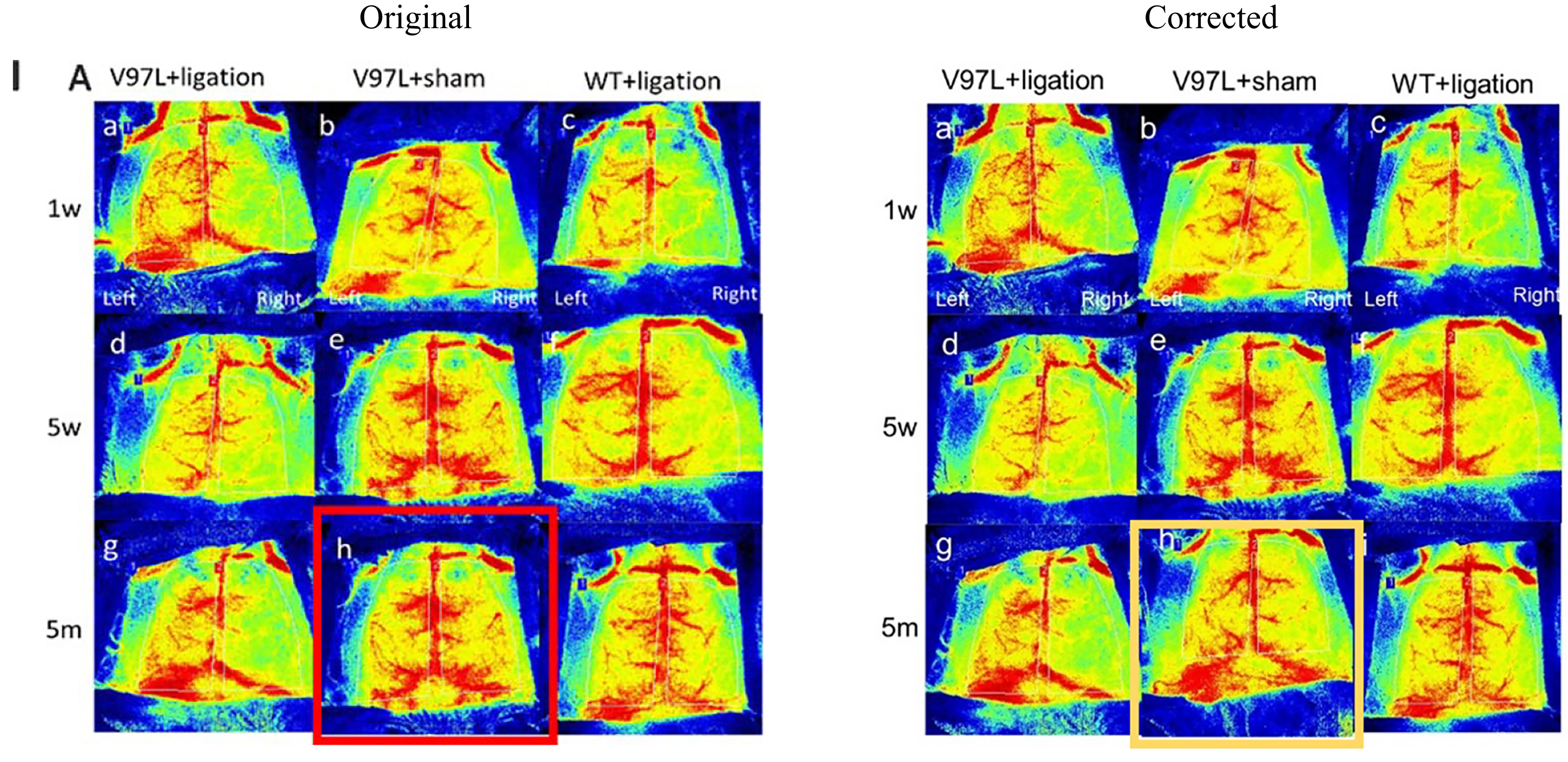
On page 266, in Fig. 1, within the “Figure A h” in the part I section, an incorrect image file was used. This picture is mainly exhibited the cerebral blood flow (CBF) at a different time point after surgery. The original part is highlighted with a red frame, and the corrected image is highlighted with a yellow frame. The full original and corrected images are also included below.
This mistake did not affect the research results and conclusion of the article.
Fig. 1
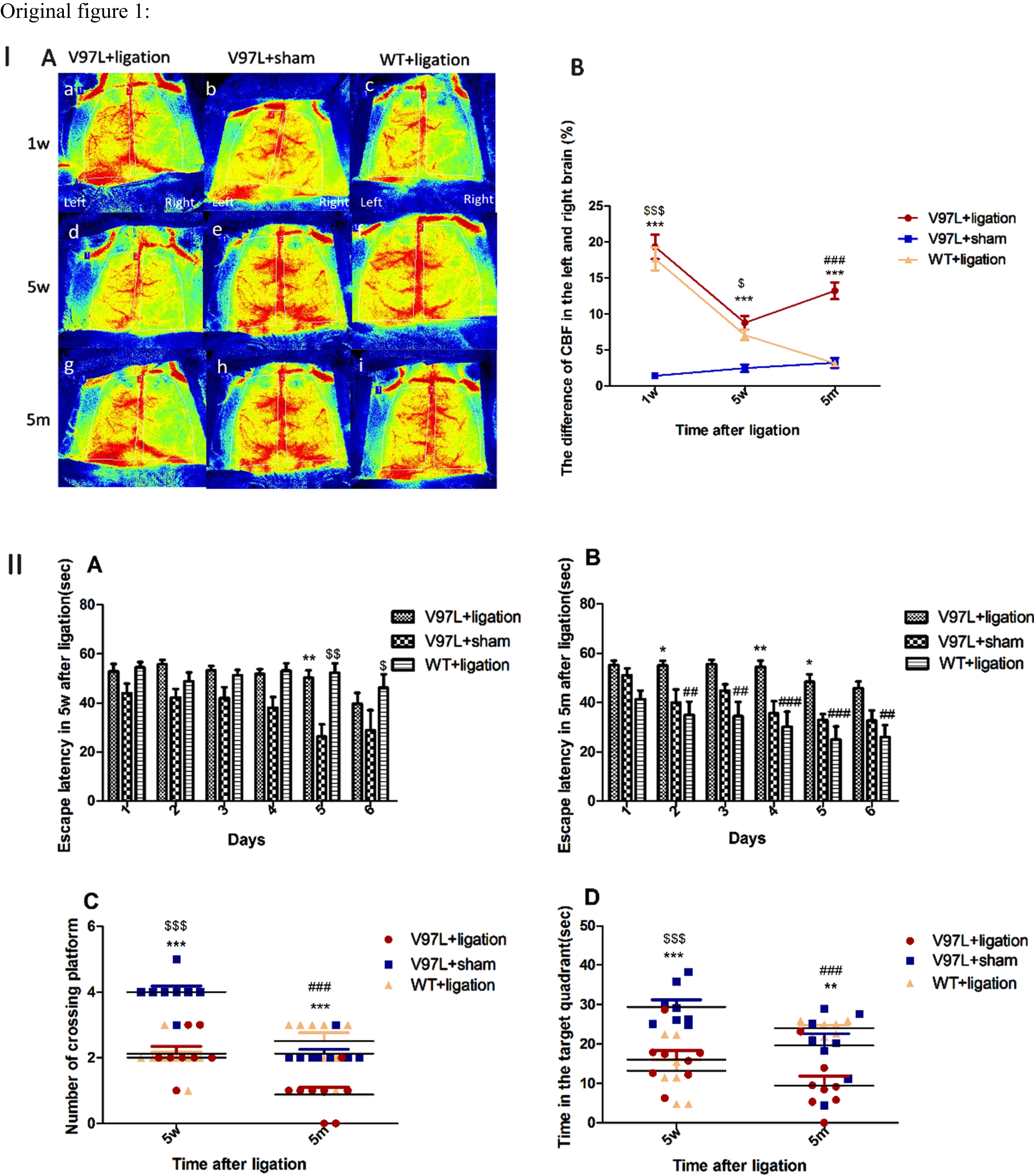
A mouse model of Alzheimer’s disease with chronic cerebral hypoperfusion was successfully developed, as indicated by disrupted cognitive function. Part I shows that (A) PS1V97L + ligation, PS1V97L + sham, and wild type (WT) + ligation mice were subjected to laser speckle blood flow monitoring to determine their cerebral blood flow (CBF) at 1 week, 5 weeks, and 5 months after surgery.
Original figure 1:
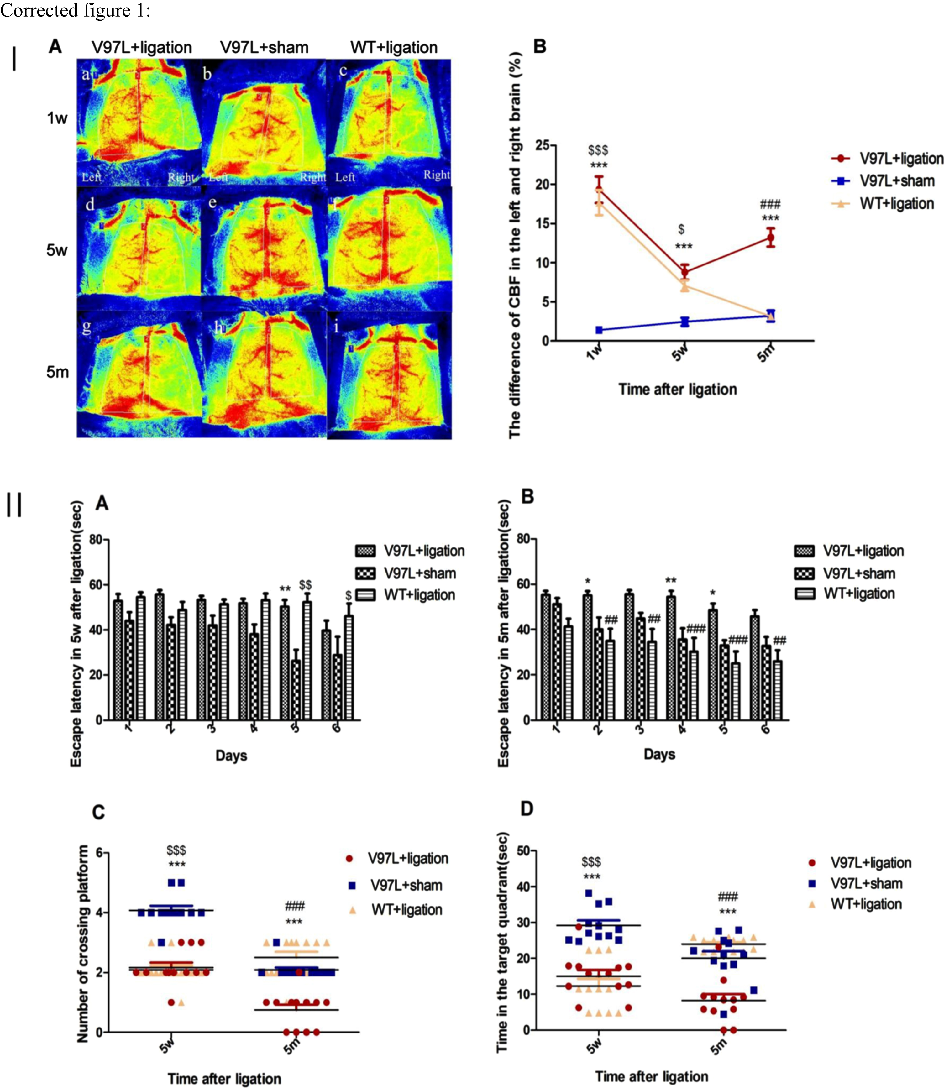
Corrected figure 1:
Fig. 1
A mouse model of Alzheimer’s disease with chronic cerebral hypoperfusion was successfully developed, as indicated by disrupted cognitive function. Part I shows that (A) PS1V97L + ligation, PS1V97L + sham, and wild type (WT) + ligation mice were subjected to laser speckle blood flow monitoring to determine their cerebral blood flow (CBF) at 1 week, 5 weeks, and 5 months after surgery. B) The difference in CBF between the left and right hemispheres of the brain. Data are shown as mean±SEM (n = 12 mice/group). Part II. A, B) Escape latency in the Morris water maze during training at 5 weeks and 5 months after surgery in PS1V97L + ligation, PS1V97L + sham, and WT+ ligation mice. C) The number of platform location crossings in the probe trial at 5 weeks and 5 months after surgery. D) The time spent in the target quadrant at 5 weeks and 5 months after surgery. Data are shown as mean±SEM (n = 12 mice/group). V97L + ligation versus V97L + sham: *p < 0.05, **p < 0.01, ***p < 0.001; V97L + ligation versus WT+ ligation: #p < 0.05, ##p < 0.01, ###p < 0.001; V97L + sham versus WT+ ligation: $p < 0.05, $$p < 0.01, $$$p < 0.001.