Erratum to: Stimulation of Dopamine Production by Sodium Benzoate, a Metabolite of Cinnamon and a Food Additive
Suresh B. Rangasamy, Sridevi Dasarathi, Aparna Nutakki, Shreya Mukherjee, Rohith Nellivalasa and Kalipada Pahan
[JAD Reports, vol. 5, no. 1, 2021; pp. 295–310, DOI: 10.3233/ADR-210001]
https://content.iospress.com/articles/journal-of-alzheimers-disease-reports/adr210001.
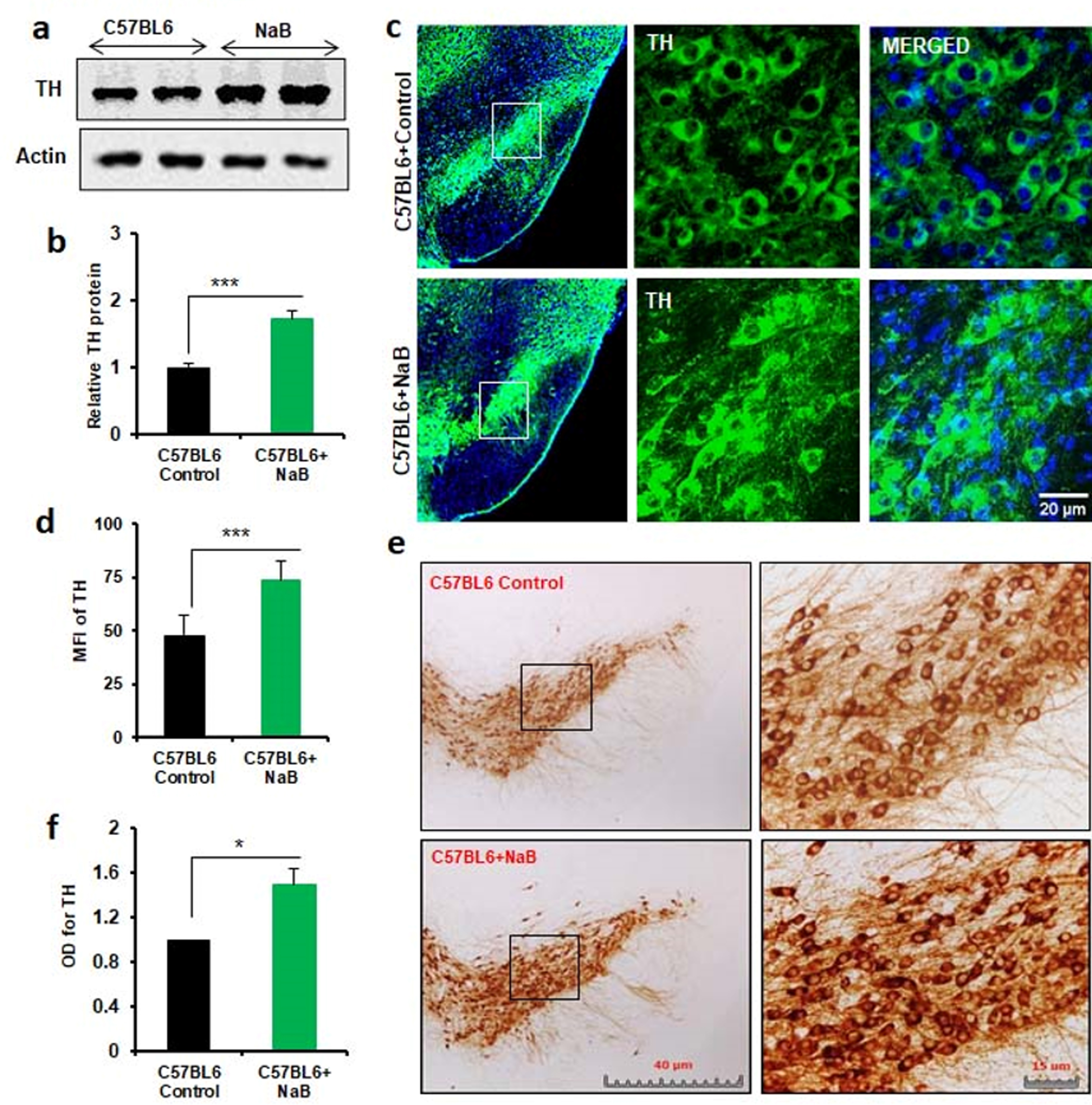
On page 304, in Figure 5C –lower panel, two incorrect images were used. This is here corrected. Please see the correct Figure 5 below.
This mistake did not affect the research results and conclusion of the article.
Fig. 5
Oral treatment with NaB increases the level of TH in vivo in the nigra of normal C57/BL6 mice. Male C57/BL6 mice (n = 5 per group) were treated with NaB (50 mg/kg body wt/d) mixed in 0.5% methylcellulose orally via gavage. Control mice received 0.5% methylcellulose as vehicle. After 30 d of treatment, the level of TH was monitored in the SNpc by Western blot (A). Actin was run as loading control. Bands were scanned and values (TH/actin) presented as relative to control (B). Results are mean±SEM of five mice per group. ***p < 0.001 vs control by two-tailed paired t-tests. The level of TH was monitored in ventral midbrain sections by immunofluorescence (C). MFI of TH (D) was calculated in two nigral sections of each of five mice per group. Results are mean±SEM of five mice per group. ***p < 0.001 vs control by two-tailed paired t-tests. The level of TH was monitored in ventral midbrain sections by DAB immunostaining (E). Optical density of TH (F) was calculated in two nigral sections of each of five mice per group. Results are mean±SEM of five mice per group. *p < 0.05 vs control by two-tailed paired t-tests.