You are viewing a javascript disabled version of the site. Please enable Javascript for this site to function properly.
Go to headerGo to navigationGo to searchGo to contentsGo to footer
In content section. Select this link to jump to navigation

H2S attenuates the myocardial fibrosis in diabetic rats through modulating PKC-ERK1/2MAPK signaling pathway

Abstract

OBJECTIVE

To investigate the roles and underlying mechanism of exogenous H2S (hydrogen sulfide) in attenuating the myocardial fibrosis in diabetic rats.

METHODS:

A total of 40 SD rats were randomly divided into 4 groups: control group, STZ group, STZ + H2S group and H2S group. To build the DM rat model , the rats in the STZ group and STZ + H2S group were injected streptozotocin (STZ) intraperitoneally, While the rats in the STZ + H2S group and the H2S group received sodium hydrosulfide (NaHS), which provides exogenous H2S. Eight weeks later, the myocardial tissues of rats were used to detecting the collagen deposition through Masson staining, as well as some protein expressions related to myocardial fibrosis and signaling pathway by western blotting.

RESULTS:

Comparing to control group, the collagen deposition of myocardial matrix remarkably increased in the STZ group, and almost all the proteins that are relative to myocardial fibrosis, inflammatory and signaling pathway show an overexpression, except for PPARG and NF-κBp65. When Compared with the STZ group, the collagen deposition was obviously attenuated in STZ + H2S group, as well as the protein expressions above-mentioned, While PPARG was up-regulated.

CONCLUSION:

The myocardial fibrosis in DM rats can be attenuated effectively by exogenous H2S, and the underlying mechanism is likely to regulating PKC-ERK1/2MAPK signaling pathway, improving the MMPs/TIMPs expression dysregulation and inhibiting inflammatory reaction.

1.Introduction

Diabetic cardiomyopathy (DCM) is one of the major chronic complications of DM, in which myocardial fibrosis is considered as the main pathological change [1]. Pathogenesis of myocardial fibrosis is quite complicated, including oxidative stress, inflammatory responses and cell apoptosis. Many studies have confirmed the close correlation between the collagen remodeling in myocardial fibrosis and dysregulation of MMPs/TIMPs expressions. Besides, as a key mediator in fibrosis [2], CTGF is involved in the fibrotic processes of multiple organs, such as kidney, liver and lung, and, according to some literatures [3, 4], CTGF also participates in the myocardial fibrosis of DM [5]. It has been found that H2S, as a new gaseous signal molecule, can exert the essential protective effect on cells in cardiovascular diseases. Some research has shown that H2S can ameliorate not only the myocardial injuries caused by ischemia reperfusion, but also the myocardial fibrosis under the overloading pressure [6], whereas the roles and relevant mechanisms of H2S in amelioration of myocardial fibrosis of DM remain unclear yet.

It has been reported in some studies that activations of TGF-β and MAPK could be induced by high glucose and non-enzymatic advanced glycation end products (AGEs) via DAG-PKC signal pathway and oxidative stress [7]. PKC, served as the hub of multiple intracellular signal pathways, is involved in the construction of key intracellular information network and regulates a variety of in-vivo physiological and pathological processes. PKC can activate the TGF-β1 which is one of the key cytokines involved in the pathogenesis [8], and some studies also reported that ERK1/2 can interact with the TGF-β1 to be involved in the fibrosis [9, 10, 11]. Thus, it can be inferred that PKC-ERK1/2MAPK may participate in the myocardial fibrosis pathogenesis in DM, or served as the underlying regulation mechanism, through which H2S can ameliorate the myocardial fibrosis in DM. In our research, DM model of rat was established by using STZ for observing the role of H2S in attenuating the myocardial fibrosis, and unveiling the regulatory effect of H2S on PKC-ERK1/2MAPK signal pathway and the underlying mechanisms to improve the expression dysregulation of MMPs/TIMPs and inflammatory responses.

2.Material and methods

2.1Experimental animals and reagents

The Animal Ethics Committee of University of South China (Hengyang, China) has approved this experimental protocol. Forty male adult SD rats (weighing 280 ± 20 g), which were purchased from the Animal Experimental Center of University of South China (Hengyang, China) and Animal qualified number is SYXK (hunan) 2015-0006,were fed in a clean laboratory with constant temperature (22 ± 1C) and artificial lighting (day and night 12 h each). Food and water were free to all rats. Streptozotocin (STZ) was provided by MP Biomedicals, LLC (USA), Sodium hydrosulfide (NaHS) was purchased from Sigma-Aldrich LLC (USA). Rabbit polyclonal antibodies against collagen I, collagen III, TGF-β1, CTGF were purchased from Bioss Biology Company (Beijing, China), rabbit polyclonal antibodies against MMP11, TIMP2, NF-κBp65, TNF-α, PPARG, HSP70, HSP90, PKCα, glyceraldehyde 3-phosphate dehydrogenase (GAPDH) and Anti-rabbit secondary antibody were all provided by Boster Biotechnology (Wuhan, China), and rabbit polyclonal antibody against ERK1/2 was obtained from Cell Signaling Technology (USA). Besides, bicinchoninic acid (BCA) Protein Assay Kit and cell lysis buffer were obtained from Beyotime Company (Shanghai, China).

2.2Preparation of models

A total of 40 male SD rats were randomly assigned to 4 groups: control group, DM group (STZ group), H2S intervention group (STZ + H2S group) and H2S-control group (H2S group). These rats were fed for 1 week to adapt to the environment. After being starved for 12 h, rats received single intraperitoneal injection of STZ (50 mg/kg; STZ being dissolved in citrate buffer; pH = 4.4) to establish the DM model of rat. For establishing the non-DM model of rat, sodium citrate buffer in the same volume was injected intraperitoneally. 72 h later, blood sample was collected from caudal veins, and those rats whose blood glucose level were higher than 16.7 mmol/L were successfully model. NaHS solution (100 μmol/kg/d) were intraperitoneally injected to the rats in STZ + H2S group and H2S group, while in control group and STZ group, NaHS solution was replaced by the normal saline in the same volume.

2.3Extraction of myocardial tissues

Eight weeks later, rats were executed immediately followed by thoracotomy to remove the heart. Heart was rinsed using normal saline to purge the residual blood, and the saline on the heart was dried using filter paper. In this study, only left ventricle and interventricular septum were preserved, from which tissues were extracted and then mounted in 10% formaldehyde for Masson staining. Remaining myocardial tissues were preserved in a -80C refrigerator.

2.4Observing the myocardial collagen deposition via Masson staining

The mounted myocardial tissues in left ventricle of rat in 4 groups were regularly embedded using paraffin and sliced into sections followed by Masson staining. Thereafter, tissues were placed under the inverted microscope for observation, in which myocardial cells were in red and collagen fibers in blue.

2.5Detecting the protein expressions of collagen-I, collagen-III, CTGF and MMPs/TIMPs in myocardial tissues via western blotting

Proteins extracted from myocardial tissues in each group were used for quantification via BCA method. Extracted proteins, after being heated for degeneration, were prepared for SDS-PAGE, and then electronically transferred onto the PVDF membrane. After the membrane was blocked using 5% BSA for 2 h, primary antibodies of collagen-I, collagen-III, CTGF, MMPs/TIMPs (diluted at 1:400), and GAPDH (internal reference; diluted at 1:1000) were added onto the membrane for incubation at 37C for 1 hour followed by incubation at 4C overnight. Thereafter, membrane was taken out and washed using TBST for 3 times. Proteins on the membrane were incubated using HRP-labelled secondary antibodies (diluted at 1:4000) at 37C for 1 h. Then, membrane was taken out and washed using TBST for 3 times. After color development using ECL, exposure and scanning, stripes on the membrane were scanned using AlphaImager software, and expression levels of each protein were presented by the ratio of gray value of targeted stripe to that of GAPDH.

2.6Detecting the protein expressions of TGF-β1, HSP70, HSP90, NF-κBp65, TNF-α and PPARG in myocardial tissues via western blotting

Samples of myocardial tissues in each group, after being sufficiently grinded, were used for protein quantification. After SDS-PAGE, the proteins were electronically transferred onto a PVDF membrane which was then incubated using the primary antibodies of TGF-β1, HSP70, HSP90, NF-κBp65, TNF-α and PPARG (diluted at 1:400) and GAPDH (internal reference; diluted at 1:1000). The rest of procedures were the same as the above.

2.7Detecting the protein expressions of PKCα and ERK1/2 in myocardial tissues via western blotting

Proteins extracted from myocardial tissues in each group were used for protein quantification. After SDS-PAGE, the proteins were electronically transferred onto a PVDF membrane which was then incubated using the primary antibodies of PKCα, and ERK1/2 (diluted at 1:400) and GAPDH (internal reference; diluted at 1:1000). The rest of procedures were the same as the above.

2.8Statistical analysis

SPSS 18.0 software was applied to analysis experimental data. Measurement data were presented as mean ± standard deviation (x¯±s). One-way ANOVA was adopted for intergroup comparison. The difference with statistical significance was presented by p< 0.05.

3.Results

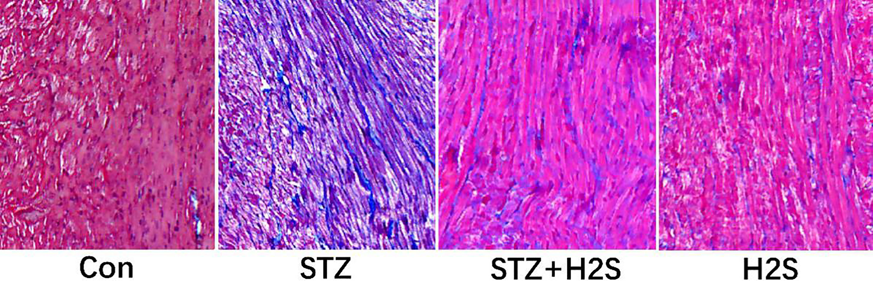
3.1Results of Masson staining

Masson staining results of myocardial tissues in left ventricle of rat in each group are shown in Fig. 1, in which the blue-stained substance is collagen fiber. Compared to control group, we observed that for rats in the STZ group, myocardium was in malalignment and collagen fiber deposition in matrix obviously increased, suggesting the obvious myocardial fibrosis. However, in comparison with the STZ group, we observed that myocardial arrangement was remarkably ameliorated and the blue-stained collagen fiber deposition was significantly reduced in rats in the STZ + H2S group, suggesting the ameliorations in myocardial fibrosis.

Figure 1.

Pathological changes of myocardium assessed by Masson staining (Images were obtained at × 100 magnification).

Pathological changes of myocardium assessed by Masson staining (Images were obtained at × 100 magnification).

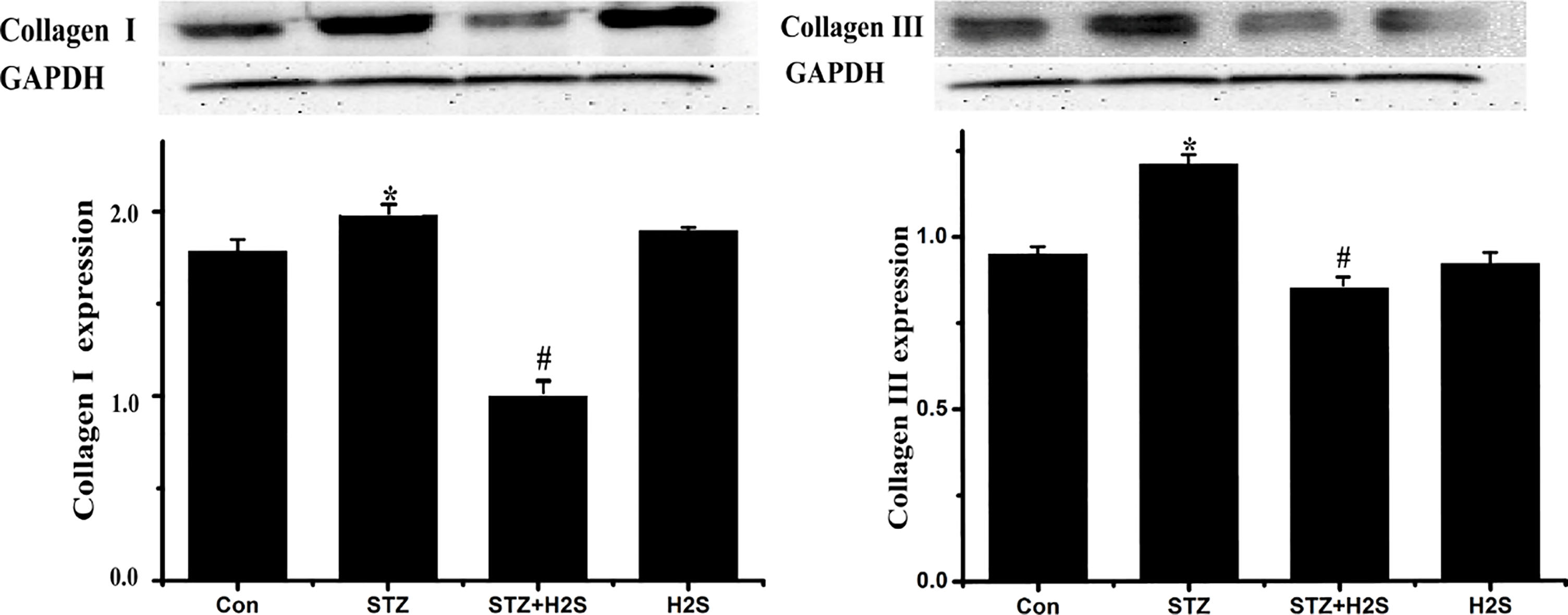
3.2Expressions of collagen-I and collagen-III in myocardial tissues of rats

As shown in Fig. 2, comparing to control group, expressions of collagen-I and collagen-III were clearly augmented (p< 0.05) in STZ group. Significant decreases were identified in STZ + H2S group when compared to STZ group (p< 0.05). Expression of collagen-I also shows difference between control group and H2S group (p< 0.05), however, the expression of collagen-III showed little variation (p> 0.05).

Figure 2.

Western blotting results of collagen-I and collagen-III. Data are expressed as mean ± SD (n= 3). P*< 0.05 vs Con group; P#< 0.05 vs STZ group.

Western blotting results of collagen-I and collagen-III. Data are expressed as mean ± SD (n= 3). P*< 0.05 vs Con group; P#< 0.05 vs STZ group.

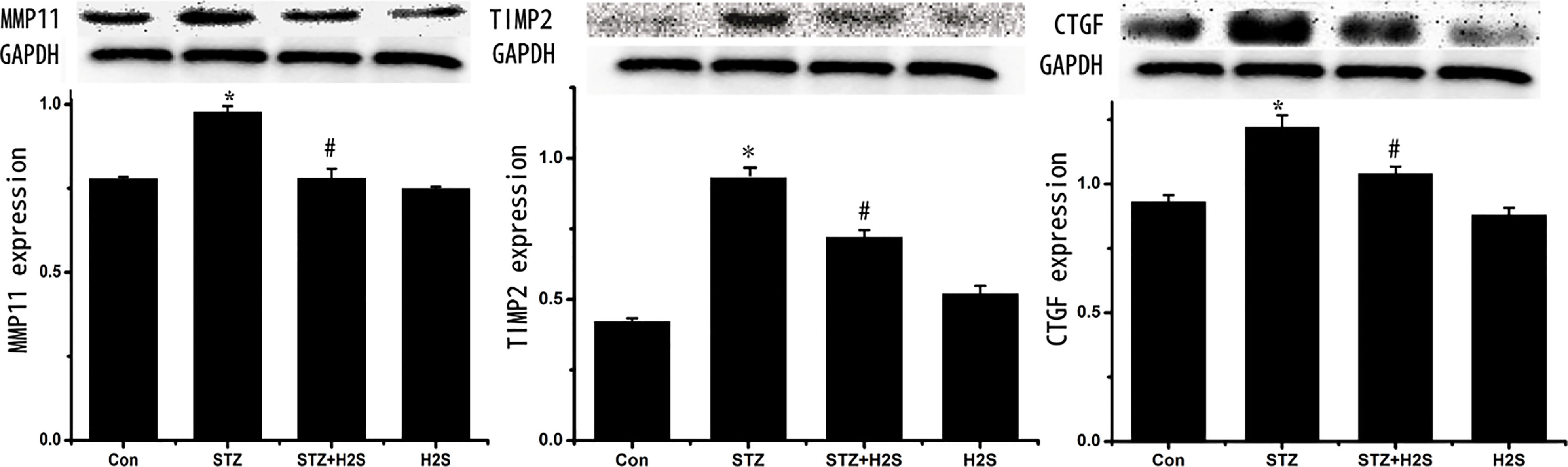
3.3Protein expressions of MMP11, TIMP2 and CTGF in myocardial tissues of rats

As shown in Fig. 3, comparing to control group, protein expressions of MMP11, TIMP2 and CTGF were significantly augmented in STZ group (p< 0.05). Significant decreases were identified in the protein expressions of rats in STZ + H2S group when comparing to STZ group (p< 0.05). However, no significant variation was detected in the protein expressions of MMP11, TIMP2 and CTGF between control group and H2S group (p> 0.05).

Figure 3.

Western blotting results of MMP11, TIMP2 and CTGF. Data are expressed as mean ± SD (n= 3). *P< 0.05 vs Con group; P#< 0.05 vs STZ group.

Western blotting results of MMP11, TIMP2 and CTGF. Data are expressed as mean ± SD (n= 3). *P< 0.05 vs Con group; P#< 0.05 vs STZ group.

3.4Protein expressions of TGF-β1, HSP70, HSP90, NF-κBp65, TNF-α and PPARG in myocardial tissues

As shown in Fig. 4, expressions of TGF-β1, HSP70, HSP90 and TNF-α were distinctly added in STZ group when compared to control group, and the PPARG was decreased (p< 0.05), while no remarkable change was observed in expression of NF-κBp65. In comparison with STZ group, significant decreases were identified in the expressions of TGF-β1, HSP70 as well as TNF-α in STZ + H2S group, while the PPARG was obviously up-regulated (p< 0.05), and no significant differences were identified in comparison of protein expressions of HSP90 and NF-κBp65 between the two groups (p> 0.05). No significative variation was presented in the protein expressions of the above genes between control group and H2S group (p> 0.05).

Figure 4.

Western blotting results of TGF-β1, HSP70, HSP90, NF-κBp65, TNF-α and PPARG. Data are expressed as mean ± SD (n= 3). *P< 0.05 vs Con group; P#< 0.05 vs STZ group, P##> 0.05 vs Con group,**P> 0.05 vs STZ group.

Western blotting results of TGF-β1, HSP70, HSP90, NF-κBp65, TNF-α and PPARG. Data are expressed as mean ± SD (n= 3). *P< 0.05 vs Con group; P#< 0.05 vs STZ group, P#⁢#> 0.05 vs Con group,**P> 0.05 vs STZ group.

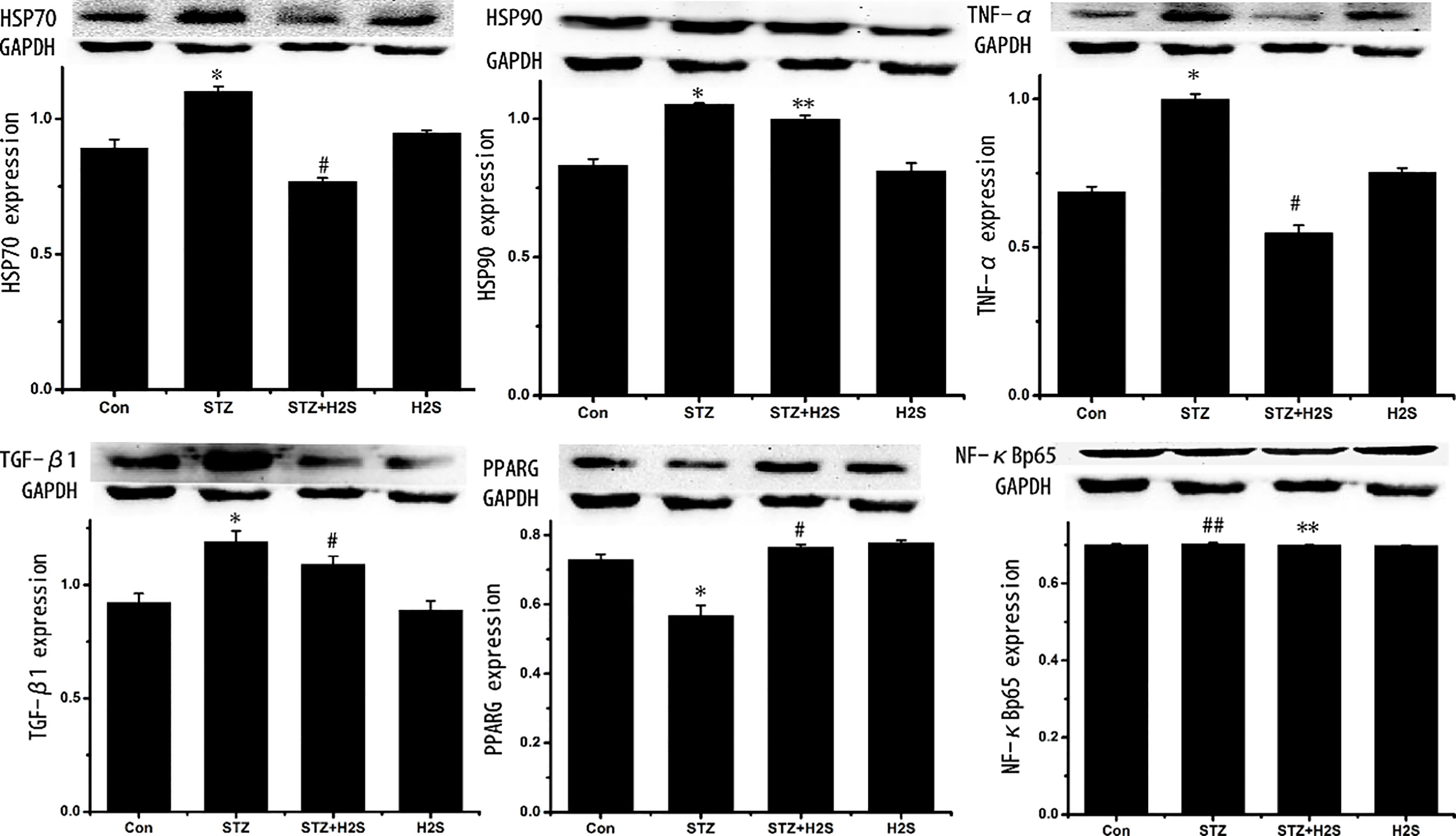
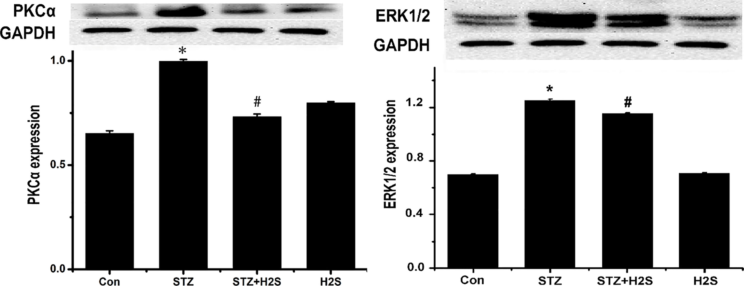
3.5Protein expressions of PKCα and ERK1/2 in myocardial tissues of rats

As shown in Fig. 5, protein expressions of PKCα and ERK1/2 were significantly augmented (p< 0.05) in STZ group when compared to control group. Compared with STZ group, significant decreases were identified in the protein expressions of PKCα and ERK1/2 in rats in STZ + H2S group (p< 0.05). However, no significant variation was detected in the protein expressions of PKCα and ERK1/2 between control group and H2S group (p> 0.05).

Figure 5.

Western blotting results of PKCα and ERK1/2. Data are expressed as mean ± SD (n= 3). *P< 0.05 vs Control group; P#< 0.05. vs STZ group.

Western blotting results of PKCα and ERK1/2. Data are expressed as mean ± SD (n= 3). *P< 0.05 vs Control group; P#< 0.05. vs STZ group.

4.Discussion

DCM is a kind of cardiovascular complications of DM, and as a kind specific cardiomyopathy, it usually has the clinical manifestations in early stage, such as left ventricular hypertrophy and decrease of diastolic function [12], and its major pathological changes include hypertrophy of myocardial cells and myocardial fibrosis. However, myocardial fibrosis is generally manifested by the excessive deposition of extracellular matrix (ECM), in which dysregulation of MMPs/TIMPs expressions is closely correlated with the deposition of ECM [13]. MMPs, a kind of Ca2+- and Zn2+-dependent proteinase, can degrade the ECM and basement membrane, while tissue inhibitors of metalloproteinase (TIMPs), as the specific inhibitor of MMPs, can suppress the activity of MMPs; thus, the balanced MMPs/TIMPs expressions are critical to maintaining the regular collagen metabolism and stability of ECM. In this study, when compared to control group, the amount of collage fibers in myocardium of rats in STZ group was significantly increased, and obvious augmentation in expressions of collagen-I and collagen-II was also observed with up-regulation in expressions of MMP11 and TIMP2, suggesting the myocardial fibrosis in DM rats. As a kind of pro-fibrosis factor that has been found in recent years, connective tissue growth factor (CTGF) can strengthen the abilities of fibroblast in mobilization, migration and ECM synthesis [14], and can be enormously expressed under the induction of TGF-β1 to participate in the pathogenesis of fibrosis in organs [15]. TGF-β1 has been widely recognized as a regulatory factor in close association with the pathogenesis of fibrosis [16], and not only can it facilitate the transformation of fibroblasts into matrix under the high glucose environment, but also it can boost the synthesis of TIMPs and suppress the degradation of newly synthesized MMPs to reduce collagen degradation and increase collagen deposition [17]. In this study, the CTGF and TGF-β1 protein expression in rats of the STZ group was significantly increased, suggested that they may be involved in the dysregulation in MMPs/TIMPs expressions and the pathogenesis of collagen remodeling in DM rats.

Myocardial fibrosis has a very complicated pathogenesis. According to the studies, inflammatory responses caused by high glucose play an important role in the myocardial fibrosis in DM, and the pro-inflammation factors induced by high glucose are critical to the initiation and progression of myocardial fibrosis [18, 19]. In these factors, TNF-α is involved in the pathogenesis and progression of myocardial fibrosis via multiple pathways, such as facilitating the proliferation of myocardial fibroblasts, inducing the oxidative stress and apoptosis of myocardial cells, enhancing the activity of MMPs and modulating the ECM synthesis. Heat shock protein 70 (HSP70) and HSP90 can reduce the degeneration of proteins under the stress status and eliminate the degenerated or misfolded polypeptides to protect the cells from damages. However, when it comes to abnormal expressions, they could induce the significant up-regulation in expression of TNF-α. Peroxisome proliferator-activated receptor γ (PPARG) can ameliorate the injuries to elastic fibers through inhibiting the inflammatory responses and protein degradation. In this study, significant myocardial fibrosis was detected in rats in STZ group with the dramatic increases in expressions of HSP70, HSP90 and TNF-α in myocardial tissues in DM rats when compared with control group, and the protein expression of PPARG was also down-regulated, indicating that inflammatory responses participate in the process of myocardial fibrosis in DM rats.

PKC-ERK1/2MAPK signal pathway is involved in the regulation mechanism of inflammatory responses and collagen remodeling in myocardial tissues in DM. PKC is a kind of phospholipid- and Ca2+-dependent protein kinase, and the activation of PKC is a key link in a series of cellular cascade signaling pathway, which can mediate the transmission of various cellular information and participate in the expression and functional modulation of multiple regulation genes. Some studies have demonstrated that PKC participates in the occurrence of myocardial hypertrophy induced by high glucose in diabetic rats [20, 21], and also can activate the TGF-β and MAPK through the DAG-PKC pathway and oxidative stress [22, 23]; MAPK is a group of highly-preserved serine/threonine kinase in cytoplasm, as one of the three main branches, ERK1/2 are somewhat more specialized for mitogenic and growth factor stimulation when activated [24], and may be served as the common pathway for vascular complications in DM and pathogenesis in target-organ damages [25, 26]. Meanwhile, both of the ERK1/2 and TGF-β1 can generate the positive feedback, in which the activated TGF-β1 can increase the expression activity, while the activated ERK1/2 can further promote the expression of TGF-β1. Pan et al. found that TGF-β1 as well as ERK1/2 were both suppressed when interstitial fibrosis and cardiac dysfunction were alleviated, suggesting that TGF-β1- ERK1/2 may be critical to the collagen remodeling [27]. In this study, the protein expressions of PKC, TGF-β1 and ERK1/2 in the myocardial tissues of rats in the STZ group were significantly up-regulated, revealing that the activation of PKC-ERK1/2MAPK might be associated with the pathological process of myocardial fibrosis in DM.

It has been found that H2S, the 3rd newly found endogenous gaseous signal molecule, can protect the heart in a variety of cardiovascular diseases (e.g. the myocardial ischemia reperfusion injuries) through pathways, such as vascular dilation, anti-inflammation, anti-oxidative stress and anti-apoptosis [28]. There are studies reporting that H2S can ameliorate the left ventricular remodeling in hypertension rats [29]; El-Seweidy et al. also found that H2S can improve the myocardial fibrosis [30]. In this study, results suggested that after the intervention of exogenous H2S, deposition of collagen fibers in myocardial matrix of DM rats, the protein expression of CTGF as well as collagen-I and collagen-II were all down-regulated, while the dysregulation in MMPs/TIMPs expressions was abated, prompting that H2S can ameliorate the myocardial fibrosis in DM rats. Additionally, our study also presented that comparing to STZ group, the protein expressions of PKCα and ERK1/2 in myocardium of rats in STZ + H2S group were down-regulated with the decrease in expressions of HSP70 and TNF-α and the increase in protein expression of PPARG, suggesting that H2S may inhibit the inflammatory responses through down-regulating the activities of PKC-ERK1/2MAPK signaling pathway, thus ameliorating the collagen remodeling in myocardial tissues in DM rats. There are also literatures reporting that endogenous or exogenous H2S in an appropriate concentration could suppress the inflammatory responses and oxidative stress through regulating the PKCα and extracellular regulated protein kinases to abate the myocardial injuries caused by ischemia or hypoxia and the complications, such as myocardial hypertrophy and fibrosis. In this study, we further confirmed that the mechanism of amelioration in myocardial collagen remodeling in DM via H2S might be associated with the regulation of PKC-ERK1/2MAPK signaling pathway. We all know DM is a chronic disease, and the myocardial fibrosis will evolve as time pass by, our research has provided new ideas for intervention in myocardial fibrosis in DM in short term, but more studies will be needed to investigate whether H2S still has positive effects as well as the specific regulation mechanism.

Acknowledgments

This study was funded by the Hunan Natural Science Foundation of China (Grant no. 2017JJ3271; 2017JJ3278).

Conflict of interest

None to report.

References

[1] 

Aneja A, Tang WH, Bansilal S, et al., Diabetic cardiomyopathy: insights into pathogenesis, diagnostic challenges, and therapeutic options. AM J Med. (2008) ; 121: (9): 748-757.

[2] 

Sakharova OV, Taal MW, Brenner BM. Pathogenesis of diabetic nephropathy: focus on transforming growth factor-beta and connective tissue growth factor. Curr Opin Nephrol Hy. (2001) ; 10: (6): 727-738.

[3] 

Gressner OA, Fang M, Li H, et al., Connective tissue growth factor (CTGF/CCN2) in serum is an indicator of fibrogenic progression and malignant transformation in patients with chronic hepatitis B infection. Clin Chim Acta. (2013) ; 5: (421): 126-131.

[4] 

Ponticos M, Holmes AM, Shi-wen X, et al., Pivotal role of connective tissue growth factor in lung fibrosis: MAPK-dependent transcriptional activation of type I collagen. Semin Arthritis Rheu. (2009) ; 60: (7): 2142-2155.

[5] 

Wang L, Yuan T, Du G, et al., The impact of 1,25-dihydroxyvitamin D3 on the expression of connective tissue growth factor and transforming growth factor-beta1 in the myocardium of rats with diabetes. Diabetes Res Clin Pr. (2014) ; 104: (2): 226-233.

[6] 

Xiao T, Luo J, Wu Z, et al., Effects of hydrogen sulfide on myocardial fibrosis and PI3K/AKT1-regulated autophagy in diabetic rats. Mol Med Rep. (2016) ; 13: : 1765-1773.

[7] 

Corderio MF. Beyond mitomycin: TGF – β and wound healing. Prog Reyin Eye Res. (2002) ; 21: (1): 75-89.

[8] 

Wu D, Peng F, Zhang B, et al., PKC-beta1 mediates glucose-induced Akt activation and TGF-beta1 upregulation in esangial cells. J Am Soc Nephrol. (2009) ; 20: (3): 554-66.

[9] 

Liu X., Gao S, Xu H, et al., lncRNAPCAT29; inhibits pulmonary fibrosis via the TGFβ1regulated RASAL1/ERK1/2 signal pathway. Molecular Medicine Reports. (2018) ; 7781-7788.

[10] 

Li X, Wang G, Qili M, et al., Aspirin Reduces Cardiac Interstitial Fibrosis by Inhibiting Erk1/2-Serpine2 and P-Akt Signalling Pathways. Cellular Physiology & Biochemistry. (2018) ; 45: (5): 1955-1965.

[11] 

Cheng M, Wu G, Song Y, et al. Celastrol-Induced Suppression of the MiR-21/ERK Signalling Pathway Attenuates Cardiac Fibrosis and Dysfunction. Cellular Physiology & Biochemistry International Journal of Experimental Cellular Physiology Biochemistry & Pharmacology. (2016) ; 38: (5): 1928-1938.

[12] 

Niemann M, Herrmann S, Ertl G, et al., Echocardiography in diabetic Cardiomyopathy. Herz. (2013) ; 38: (1): 42-47.

[13] 

Alameddine HS, Morgan JE. Matrix Metalloproteinases and Tissue Inhibitor of Metalloproteinases in Inflammation and Fibrosis of Skeletal Muscles. Journal of Neuromuscular Diseases. (2016) ; 3: (4): 455-473.

[14] 

Lipson KE, Wong C, Teng Y, et al., CTGF is a central mediator of tissue remodeling and fibrosis and its inhibition can reverse the process of fibrosis. Fibrogenesis & Tissue Repair. (2012) ; 5: (Suppl 1): S24.

[15] 

Igarashi A, Okochi H, Bradham DM, et al., Regulation of connective tissue growth factor gene expression in human skin fibroblasts and during wound repair. Mol Biol Cell. (1993) ; 4: (6): 637-645.

[16] 

Huang P, Zhang YJ, Huang Y, et al., Effect of PKC signalling pathway and aldose reductase on expression of fibronectin induced by transforming growth factor-β1 in human mesangial cells. Zhonghua Bing Li Xue Za Zhi. (2010) ; 39: (6): 405-409.

[17] 

Gomes LR, Terra LF, Wailemann RA, et al., TGF-β1 modulates the homeostasis between MMPs and MMP inhibitors through p38 MAPK and ERK1/2 in highly invasive breast cancer cells. BMC Cancer. (2012) ; 12: : 26.

[18] 

Malfitano C, Alba-Loureiro TC, Rodrigues B, et al., Hyperglycaemia protects the heart after myocardial infarction: aspects of programmed cell survival and cell death. Eur J Heart Fail. (2010) ; 12: (7): 659-667.

[19] 

Sheng J, Shim W, Wei H, et al., Hydrogen sulfide suppresses human atrial fibroblast proliferation and transformation to myofibroblastss. J Cell Mol Med. (2013) ; 17: (10): 1345-1354.

[20] 

Yan L, Huang H, Tang QZ, et al., Breviscapine protects against cardiac hypertrophy through blocking PKC-alpha-dependent signaling. J Cell Biochem. (2010) ; 109: : 1158-71.

[21] 

Wang M, Zhang WB, Zhu JH, Fu GS, Zhou BQ. Breviscapine ameliorates hypertrophy of cardiomyocytes induced by high glucose in diabetic rats via the PKC signaling pathway. Acta Pharmacol Sin. (2009) ; 30: : 1081-91.

[22] 

Ryu JM, Lee MY, Yun SP, et al., High glucose regulates cyclin D1/E of human mesenchymal stem cells through TGF-beta1 expression via Ca2+/PKC/MAPKs and PI3K/Akt/mTOR signal pathways. J Cell Physiol. (2010) ; 224: (1): 59-70.

[23] 

Igarashi M, Wakasak H, Takahara N, et al., Glucose or diabetes activates p38MAPK via different pathways J. Chin Invest. (1999) ; 103: (2): 185-195.

[24] 

Kehat I, Molkentin JD. Extracellular Signal-Regulated Kinases 1/2 (ERK1/2) Signaling in Cardiac Hypertrophy. Annals of the New York Academy of Sciences. (2010) ; 96-102.

[25] 

He M, Xue Z, Li J, et al., Breviscapine inhibits high glucose-induced proliferation and migration of cultured vascular smooth muscle cells of rats via suppressing the ERK1/2 MAPK signaling pathway. Acta Pharmacol Sin. (2012) ; 33: (5): 606-614.

[26] 

Panchatcharam M, Miriyala S, Yang F, Leitges M, Chrzanowska-Wodnicka M, Quilliam LA, et al., Enhanced proliferation and migration of vascular smooth muscle cells in response to vascular injury under hyperglycemic conditions is controlled by beta3 integrin signaling. Int J Biochem Cell Biol. (2010) ; 42: : 965-74.

[27] 

Pan Z, Zhao W, Zhang X, Wang B, Wang J, Sun X, et al., Scutellarin alleviates interstitial fibrosis and cardiac dysfunction of infarct rats by inhibiting TGFbeta1 expression and activation of p38-MAPK and ERK1/2. Br J Pharmacol. (2011) ; 162: : 688-700.

[28] 

Xu W, Chen J, Lin J, et al., Exogenous H2S protects H9c2 cardiac cells against high glucose-induced injury and inflammation by inhibiting the activation of the NF-κB and IL-1β pathways. Int J Mol Med. (2015) ; 35: : 177-186.

[29] 

Kondo K, Bhushan S, King AL, et al., H2S Protects Against Pressure Overload Induced Heart Failure via Upregulation of Endothelial Nitric Oxide Synthase (eNOS). Circulation. (2013) ; 127: (10): 1116-1127.

[30] 

El-Seweidy MM, Sadik NA, Shaker OG. Role of sulfurous mineral water and sodium hydrosulfide as potent inhibitors of fibrosis in the heart of diabetic rats. Arch Biochem Biophys. (2011) ; 506: (1): 48-57.