
Ectopic insertion of a duplicated ureter into prostatic urethra: Demonstration by 3D multi-detector computed tomography urography
Abstract
Ectopic insertion of the ureter in the genitourinary tract is a rare congenital disorder, usually associated with ureteral duplication. Identification of the insertion open is critical for ureteric re-implantation. However, the challenge in the diagnosis of ectopic insertion of the ureter usually is to identify its insertion, particularly when the affected ureter is not dilated. Multi-detector computed tomography (MDCT) urography with nonionic iodinated contrast media delineates the ureteric course in the normal functioning kidney in the excretory phase [1]. This report presented a young male patient with ectopic insertion of a duplicated ureter diagnosed by MDCT urography. Three-dimensional (3D) analysis technology, such as volume rendering (with a color display improving the visualization of complex anatomy and 3D relationships) and maximum intensity projection (similar in principle to projection angiography), is useful for the illustration of urinary tract anatomy [1]. Rotated volume rendering reconstruction images and continual thinner maximum intensity projection reformatted images can be viewed as videos, which provides detail delineation of the ectopic ureteral insertion and its associated ureteral duplication.
In this study, we reported MDCT urography and 3D analysis technology as an appropriate diagnostic method for the ectopic ureteral insertion and its associated complications.
1Introduction
Ectopic insertion of the ureter is defined as abnormal insertion of the ureter [2]. The incidence of ectopic ureter is about 1 : 2000 [3], and it occurs more frequently in females than in males at a ratio of approximately 6 : 1 [3–5]. In women, the ectopic orifice is either suprasphincterically or infrasphincterically, thus the ectopic insertion sites may locate at lower bladder, urethra, vestibule, vagina, uterus, or Gartner duct cyst [5, 6]. In men, the extravesical ectopic orifice is always suprasphincterically and the ectopic orifice sites may be in bladder neck, seminal vesicle, vas deferens, prostate or epididymis [7]. Normal urination together with continuous incontinence are patognomic features for infrasphincteric ureteral openings [8].
Ectopic ureteral insertion is most commonly associated with complete duplication and obstruction [2]. The challenge in the diagnosis of ectopic insertion of the ureter is to find its insertion opening [7]. MDCT urography with 3D imaging techniques can improve temporal and spatial resolutions of the latest CT scanners and be able to acquire isotropic datasets with identical resolutions in all three planes. It provides results that are equivalent, and almost certainly superior, to those of conventional excretory urography in the evaluation of the upper urinary tracts for transitional cell carcinoma [1]. Herein, we present a young male patient with ectopic insertion of a duplicated ureter was diagnosed by MDCT urography.
2Case report
A 27-year-old male patient presented with intermittent lower left flank pain over 3 years. Routine urine and blood examinations were within normal limits. Initially, ultrasonography (USG) demonstrated double collecting system in left kidney, and moderate hydroureteronephrosis of the collecting system draining upper pole of the left kidney. MDCT urography demonstrated the ureter draining the upper pole of the kidney was dilated (Fig. 1a) and opened into the prostatic urethra (Fig. 1b and c), while other ureter draining the lower pole of the left kidney was normal. As the upper pole of the left kidney remaining function, ureteric re-implantation on the left posterior bladder wall was performed for the ectopic ureter and its distal stump was ligated. The patient’s postoperative course was uneventful and his symptoms resolved completely.
3Discussion
Ectopic insertion of the ureter in the genitourinary tract is often accompanied with ureteral duplication [2]. Partial duplication of the collecting system results from the branching of the ureteral bud before it connects with the metanephric blastemal [9]. Complete duplication occurs when two separate ureteric buds arise from the mesonephric duct. According to Weigert-Meyer law, the upper-moiety ureter usually inserts into the inferior and medial portion of the urinary bladder, while the lower-moiety ureter usually inserts into the bladder trigone [9].
Several imaging modalities are available in diagnosis of the congenital defect in the genitourinary tract system. Each of the methods has its own advantages and disadvantages (Table 1). 99mTc-DMSA scintigraphy is mainly used for the functional study of choice in identifying poorly functioning renal tissue [10]. Magnetic resonance imaging offers dilated collecting system, ectopic ureter and its extravesical insertion point [6]. Most of the partial duplications and uncomplicated complete duplication are incidentally detected on USG or intravenous urography (IVU), while ectopic ureteral insertion sites may not be easily visible [4, 11]. The early diagnosis of potential complications of this anomaly should be made in order to prevent permanent renal damage. Therefore, sufficient information about ectopic ureteral orifice site (suprasphincterical or infrasphincterical levels) and complications (with or without hydronephrosis) could be obtained by MDCT urography [11].
MDCT urography utilizes 3D analysis technology along with the administration of intravenous contrast to create images similar those obtained with IVU [11, 12]. Volume rendering is the visualization and manipulation of objects represented as sampled data in three or more dimensions [1]. The maximum intensity projection technique displays the pixels of greatest intensity along a predefined axis of the image [1, 13]. The detail information is useful for the depiction of urinary tract anatomy when there is a large difference between attenuation values of urinary tract opacified by contrast agent, and the surrounding tissues [12, 14]. Rotated volume rendering reconstruction images and continual thinner maximum intensity projection reformatted images can be observed in the videos, and can offer good delineation of the ectopic ureteral orifice and its associated ureteral duplication.
The surgical management of ectopic ureter aims at preserving renal function, eliminating infection, and maintaining continence [10]. In a functioning upper pole, the procedures that can be done are distal and proximal uretero-ureterostomy (end-to-side), or upper polar ureteric re-implantation. In cases of nonfunctional moiety, an upper polar nephrectomy may be performed to prevent the risk of infection. In this case, the upper polar renal function remaining was considered sufficient to justify ureteric re-implantation.
References
[1] | Raman S.P. , Horton K.M. and Fishman E.K. , MDCT evaluation of ureteral tumors: Advantages of 3D reconstruction and volume visualization, AJR Am J Roentgenol 201: ((2013) ), 1239–1247. |
[2] | Senel U. , Tanriverdi H.I. , Ozmen Z. and Sozubir S. , Ectopic ureter accompanied by duplicated ureter: Three cases, J Clin Diagn Res 9: ((2015) ), PD2–PD10. |
[3] | Dudek-Warchoł T. , Szmigielska A. , Krzemien G. and Warchoł S. , Ectopic ureter, renal dysplasia, and recurrent epididymitis in an infant: Case report and review of the literature, Clin Case Rep 2: ((2014) ), 7–9. |
[4] | Lawler L.P. , Jarret T.W. , Corl F.M. and Fishman E.K. , Adult ureteropelvic junction obstruction: Insights with three-dimensional multi-detector row CT, Radiographics 25: ((2005) ), 121–134. |
[5] | McNab A. and Williams C. , Perioperative anuria resulting in the diagnosis of infrasphincteric ectopic ureter in an adult female, Urol Case Rep 2: ((2014) ), 121–122. |
[6] | Demir M. , Çiftçi H. , Kiliçarslan N., Gümüs K., Oğur M., Gülüm M. and Yeni E., A case of an ectopic ureter with vaginal insertion diagnosed in adulthood, Turk J Urol 41: ((2015) ), 53–55. |
[7] | El-Ghar M.A. and El-Diasty T. , Ectopic insertion of the ureter into the seminal vesicle, World J Radiol 5: ((2013) ), 349–351. |
[8] | Carrico C. and Lebowitz R.L. , Incontinence due to an infrasphincteric ectopic ureter: Why the delay in diagnosis and what the radiologist can do about it, Pediatr Radiol 28: ((1998) ), 942–949. |
[9] | Fernbach S.K. , Feinstein K.A. , Spencer K. and Lindstrom C.A. , Ureteral duplication and its complications, Radiographics 17: ((1997) ), 109–127. |
[10] | Ghosh B. , Shridhar K. , Pal D.K. and Banerjee M. , Ectopic ureter draining into the uterus, Urol Ann 8: ((2016) ), 105–107. |
[11] | Hu X.Y. , Hu C.H. , Fang X.M. , Yao X.J. , Lerner A. , Chen H.W. and Zhu Z.M. , Practical value of intravenous urography combined with add-on CT in diagnosing ureteral abnormalities, Chin Med J (Engl) 125: ((2012) ), 1287–1291. |
[12] | Song L. , Xie M. , Zhang Y. and Xu Y. , Imaging techniques for the diagnosis of male traumatic urethral strictures, J Xray Sci Technol 21: ((2013) ), 111–123. |
[13] | Lu X. , Wu R. , Huang X. and Zhang Y. , Noncontrast multidetector-row computed tomography scanning for detection of radiolucent calculi in acute renal insufficiency caused by bilateral ureteral obstruction of ceftriaxone crystals, J Xray Sci Technol 20: ((2012) ), 11–16. |
[14] | Sa Y.L. , Xu Y.M. , Feng C. , Ye X.X. and Song L.J. , Three-dimensional spiral computed tomographic cysto-urethrography for post-traumatic complex posterior urethral strictures associated with urethral-rectal fistula, J Xray Sci Technol 21: ((2013) ), 133–139. |
Figures and Tables
Fig.1
MDCT urography 3D analysis on complete duplication of the left collecting system with ectopic ureteral insertion. (a), Rotated volume rendering reconstruction images show complete duplication of the collecting system in the left kidney, and mild hydronephrosis of upper moiety. (b, c), Continual thinner coronal maximum intensity projection reformatted images show the ureter draining the upper moiety dilated and opening into the prostatic urethra (short arrow), the other ureter draining the lower moiety normal and opening into the left posterolateral aspect of the bladder trigone (longarrow).
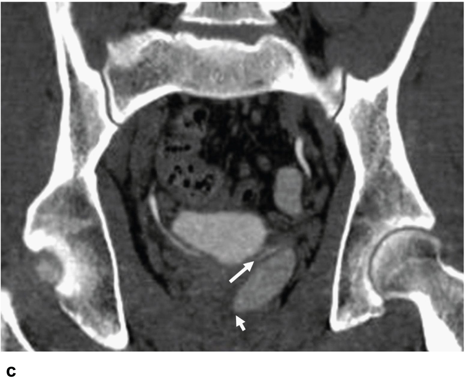
Table 1
Advantages and disadvantages of imaging modalities for ectopic ureteral insertion
| Imaging modalities | Advantages | Disadvantages |
| 99mTc-DMSA scintigraphy | –Evaluating renal function | –Ionizing radiation |
| Ultrasonography | –Quick and inexpensive | –Ectopic ureter with dysplastic kidney may not be visualized |
| –No ionizing radiation | –Inefficient in detecting ectopic ureteral orifice | |
| Intravenous urography | –Gathering sufficient information about the ectopic ureteral insertion | –Ectopic ureter with dysplastic kidney may not be visualized |
| –Ionizing radiation | ||
| –Iodinated contrast medium side effect | ||
| Computed tomography | –Providing good delineation of the ectopic ureter and its insertion | –Ionizing radiation |
| urography | –Reformatted images can be viewed as videos with 3D analysis technology | –Iodinated contrast medium sideeffect |
| Magnetic resonance | –No ionizing radiation | –Time consuming |
| Imaging | –Expensive |


