Erratum to: Tubastatin A/ACY-1215 Improves Cognition in Alzheimer’s Disease Transgenic Mice
Ling Zhang, Cui Liu, Jie Wu, Jing-jing Tao, Xiao-long Sui, Zhi-gang Yao, Yan-feng Xu, Lan Huang, Hua Zhu, Shu-li Sheng and Chuan Qin
[Journal of Alzheimer’s Disease, 41(4) (2014) 1193-1205, DOI 10.3233/JAD-140066]
http://doi.org/10.3233/JAD-140066
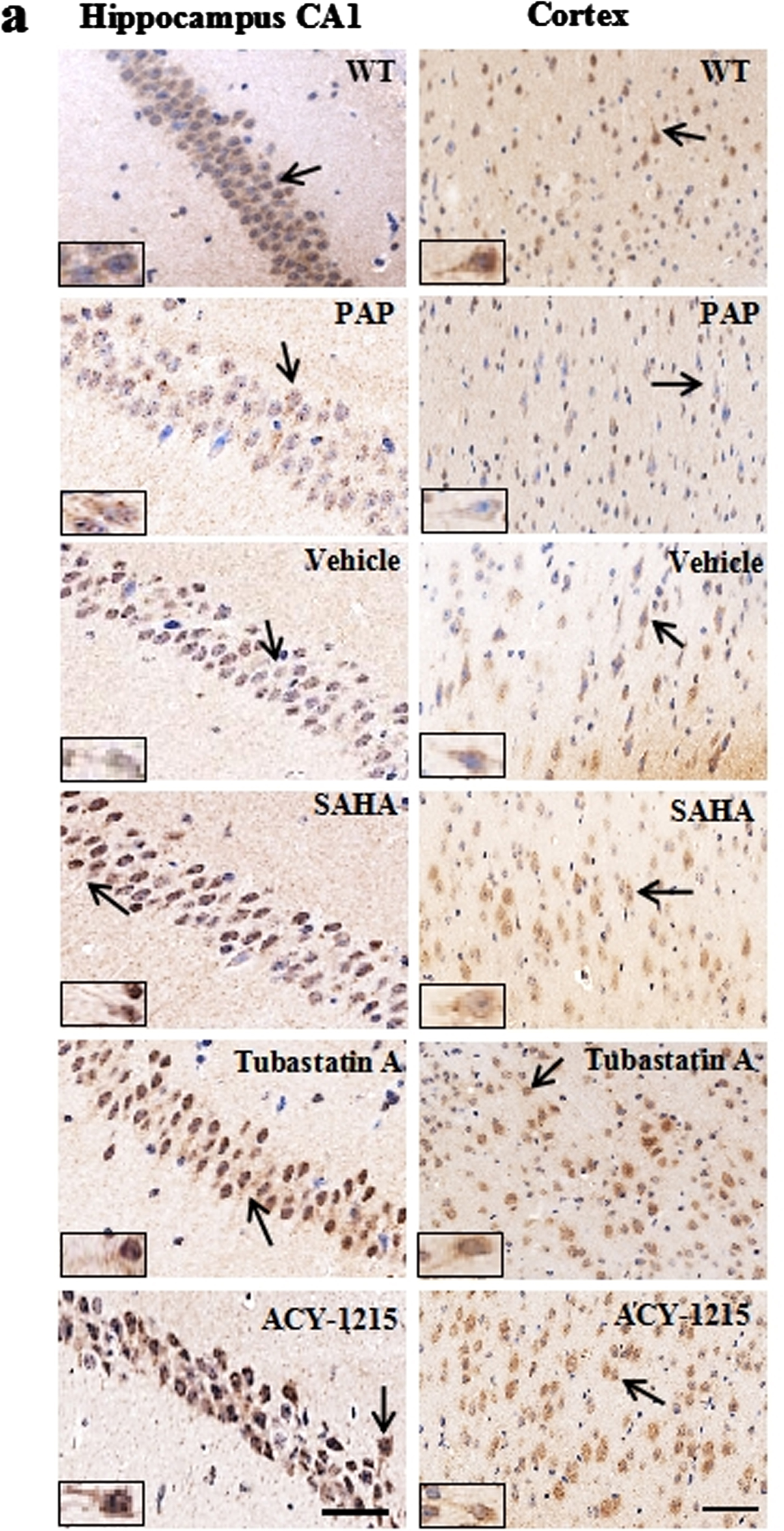
On page 1197, a pathological picture of WT was misplaced (left, first row). The wrong one was replaced in this corrected Fig. 2a.
Fig. 2
(a). Representative images showing increased immunoreactivity of ace-α-tubulin (K40) in the hippocampus (left) and prefrontal cortex (right) after Tubastatin A or ACY-1215 treatment. Scale bar: 50 μm.
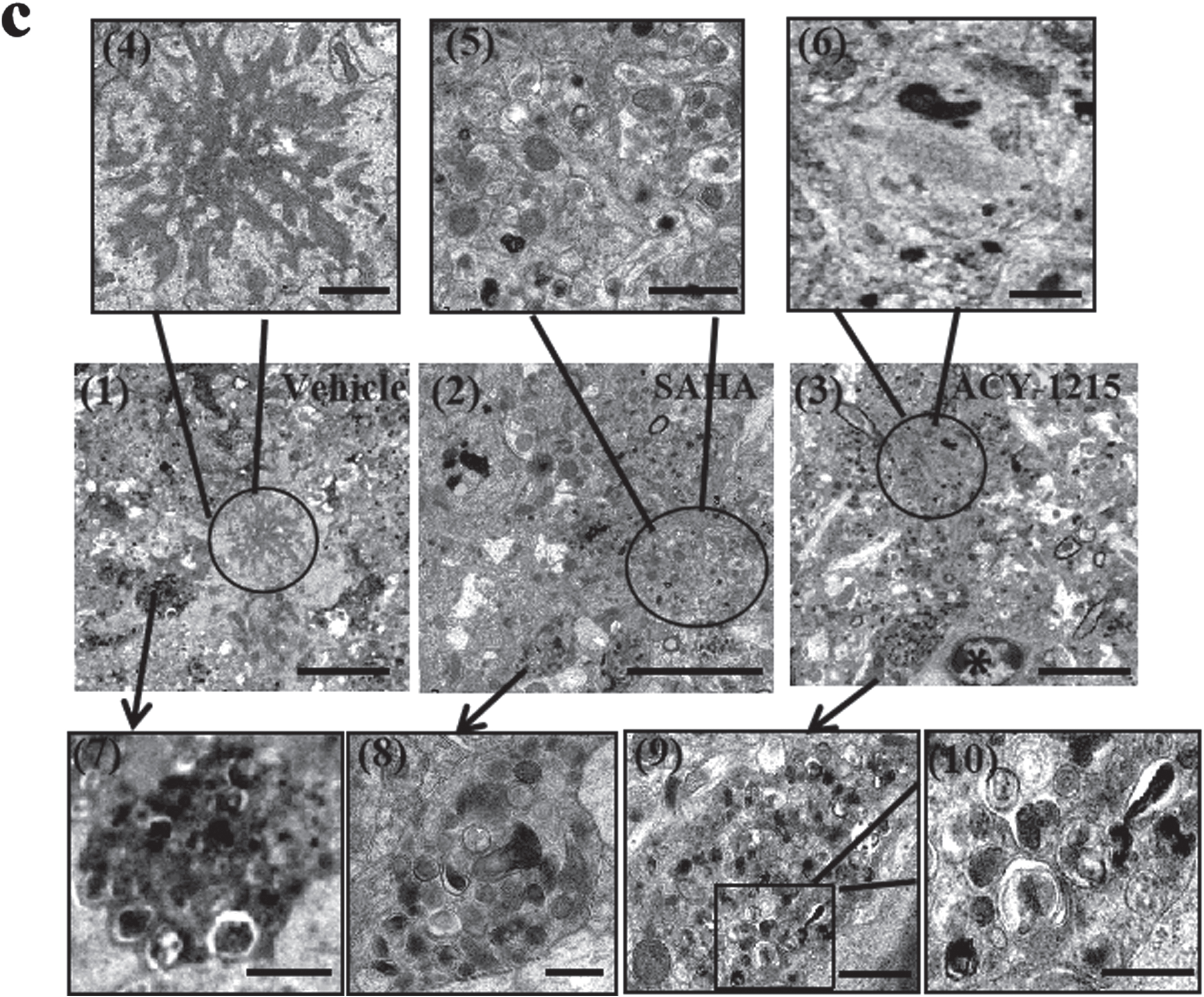
On page 1198, a picture of SAHA was misplaced (middle). The wrong ones were replaced in this corrected Fig. 3c.
Fig. 3
(c) Representative electron microscopy of senile plaques surrounded by dystrophic neurites, autophagic vesicles (AVs) and microglia (asterisk) in pre-frontal cortex of the three groups (vehicle, SAHA and ACY-1215). Flamelike plaque cores, present in the vehicle-treated group (1, 4), was absent in the HDAC6i-treated groups (2, 3, 5, 6). The number of AVs was reduced (7–10) and axon degeneration (2, 3) was improved in the HDACi-treated groups (n = 3). Scale bar: 5 μm (1, 2, 3); 1 μm (4–9); 500 nm (10).